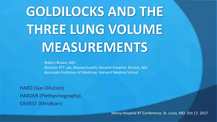
o HARDER (Plethysmography) o EASIEST (MiniBox+) Mercy Hospital RT - - PowerPoint PPT Presentation

o harder plethysmography
SMART_READER_LITE
LIVE PREVIEW

o HARDER (Plethysmography) o EASIEST (MiniBox+) Mercy Hospital RT - - PowerPoint PPT Presentation

Robert Brown, MD Director PFT Lab, Massachusetts General Hospital, Boston, MA Associate Professor of Medicine, Harvard Medical School o HARD (Gas Dilution) o HARDER (Plethysmography) o EASIEST (MiniBox+) Mercy Hospital RT Conference, St. Louis,


slide-1
SLIDE 1
  • HARD (Gas Dilution)
  • HARDER (Plethysmography)
  • EASIEST (MiniBox+)

Robert Brown, MD Director PFT Lab, Massachusetts General Hospital, Boston, MA Associate Professor of Medicine, Harvard Medical School Mercy Hospital RT Conference, St. Louis, MO Oct 17, 2017

slide-2
SLIDE 2

 NONE  ON SAB OF PULM-ONE  NO PAYMENT, NO EQUITY

Disclosure

slide-3
SLIDE 3
slide-4
SLIDE 4
slide-5
SLIDE 5
slide-6
SLIDE 6
slide-7
SLIDE 7
slide-8
SLIDE 8

 Solution in H2O (negligible)  Equipment leaks (equilibration fails)  Leaks around nose clip, mouthpiece  Ruptured tympanic membrane  Swallowing  Absorption into fluids, tissues

Paths of Helium Loss

slide-9
SLIDE 9
slide-10
SLIDE 10

 Range 0-10%  Resolution: 0.01%  95% response time of < 15 sec to 2% step change in HE

concentration

 < 0.02% drift for 10 minutes

HELIUM ANALYZER

slide-11
SLIDE 11

 Δ [HE] < 0.02% in 30 secs (3x, z 15 seconds)  Based on:

 Limits of resolution of He meter  Estimate of Δ [He] of < 0.01% per minute due to He absorption and

N2 excretion

End of Test Criteria

slide-12
SLIDE 12
slide-13
SLIDE 13
slide-14
SLIDE 14
slide-15
SLIDE 15
slide-16
SLIDE 16
slide-17
SLIDE 17

 NORMAL: results very similar  RESTRICTION: results very similar  OBSTRUCTION: Pleth>helium but……CONTROVERSIAL

Helium vs Plethysmograph

slide-18
SLIDE 18

 REFERENCE:

 Comparison of Plethysmograph and Helium Dilution

Lung Volumes: which is best in COPD? O’Donnell CR, Brown R, Loring SH et al CHEST 2010; 137: 1116-1121

Helium vs Plethysmograph controversy

slide-19
SLIDE 19
slide-20
SLIDE 20
slide-21
SLIDE 21 21

 Measurements

 Spirometry

○ FEV1 ○ FEV6 ○ FEF25-75%

 Lung Volume Measurement

○ TLC and subdivisions

 Diffusion

○ CO Diffusion capacity ○ Real-time analysis/rapid gas analyzer

MiniBox+

slide-22
SLIDE 22 22

 Key Features

 Easy to learn, easy to use touch screen tablet  Small footprint  Low maintenance  Low cost per test

MiniBox+

slide-23
SLIDE 23 23

 Meets ATS/ERS standards

 Heated pressure differential pneumotach  End of test criteria Progress Wheel

○ At least 6 seconds and <25 ml volume change for 1-second

Spirometry

slide-24
SLIDE 24
slide-25
SLIDE 25
slide-26
SLIDE 26
slide-27
SLIDE 27 27

 Meets ATS/ERS standards

 Multi-gas analyzer for CO, CH4  Single gas tank

○ 0.300% CO, 0.300% CH4, 21% O2, Balance N2

Diffusion

slide-28
SLIDE 28
slide-29
SLIDE 29
slide-30
SLIDE 30 30

 Traditional

 Gas dilution

○ Long duration (4-7 minutes) ○ Wait time for repeatability

 Body Plethysmography

○ Specific panting maneuver ○ No airflow for 4-7 seconds ○ Respiratory maneuver complex ○ Technologist influence

Lung Volume Measurement

slide-31
SLIDE 31 31

 MiniBox+

 Innovative solution  Tidal breathing and slow vital capacity  ~90 seconds to complete  Respiratory maneuver not complex

Lung Volume Measurement

slide-32
SLIDE 32 32

 How it works

 6 interruptions during tidal breathing

○ Rapid 100 msec shutter close and open (mouth pressure)

 Effect of interruptions on internal box

○ Pressure and flow

 Data Model

○ Proprietary Model and Algorithm

 Thoracic Gas Volume (TGV) is determined from which FRC

is measured spirometrically as the difference between TGV and end-expiratory lung volume

Lung Volume Measurement

slide-33
SLIDE 33 33

Lung Volume Measurement

  • Interruption is during inspiration (~1/3 of normal tidal)
  • Interruption counter provides feedback on when interruption segment is complete
  • Slow vital capacity can be performed either IC, VC or ERV, VC method
slide-34
SLIDE 34 34

Lung Volume Measurement

No wait time requirement between efforts No body cabin No complex panting maneuver against an

  • ccluded airway
slide-35
SLIDE 35 35

 Comparison – CT scan vs. plethysmography

Lung Volume Measurement

O’Donnell, Carl R. (2010). Comparison of Plethysmographic and Helium Dilution Lung

  • Volumes. CHEST, 137(5), 1108-1115.
slide-36
SLIDE 36 36

 Comparison – He dil. vs. plethysmography

Lung Volume Measurement

O’Donnell, Carl R. (2010). Comparison of Plethysmographic and Helium Dilution Lung

  • Volumes. CHEST, 137(5), 1108-1115.
slide-37
SLIDE 37 37

MiniBox+ Lung Volume Measurement

N=300

slide-38
SLIDE 38 38

MiniBox+ Lung Volume Measurement

 Comparison – MiniBox+ vs. Plethysmography

slide-39
SLIDE 39 39

MiniBox+ Lung Volume Measurement

Cincinnati Study Boston Study

  • Adj. R2=0.85

CV=10.9%

  • Adj. R2=0.85

CV=9.1%

slide-40
SLIDE 40 40

MiniBox+ Lung Volume Measurement

slide-41
SLIDE 41 41

 Review

 Unique and innovative technique  Compares well to Body Plethysmography  Simple respiratory maneuvers that are easy for the patient to perform  Very user friendly for technicians and patients

MiniBox+ Lung Volume Measurement

slide-42
SLIDE 42 42

 Review (cont.)

 Short test time (~90 seconds)  Multiple efforts without wait period  Small footprint  Notably less expensive than body plethysmography

MiniBox+ Lung Volume Measurement

slide-43
SLIDE 43

Q&A