Nervous System Overview functional and structural overview - - PowerPoint PPT Presentation
Nervous System Overview functional and structural overview histology electrophysiology synaptic connections neurotransmitters sensory receptors neural integration Functional overview 3 primary functions sensory
Overview
- functional and structural overview
- histology
- electrophysiology
- synaptic connections
- neurotransmitters
- sensory receptors
- neural integration
Functional overview
3 primary functions
- sensory input
- integration
- motor output
Structural overview
- Central nervous system (CNS)
- brain
- spinal cord
- Peripheral nervous system
(PNS)
- sensory
- motor
§ somatic (voluntary) § autonomic (involuntary)
- sympathetic (mobilizing)
- parasympathetic
(housekeeping)
PNS function
- Sensory (afferent) division
- Signals travel from receptors to CNS
§ Receptors - cells and organs that detect stimuli
- Motor (efferent) division
- Signals travel from CNS to effectors
§ Effectors – glands and organs that carry out the
response
Sensory Division
- Visceral sensory division
- Signals from the viscera to the CNS
§ Viscera – heart, lungs, stomach, etc.
- Somatic sensory division
- Signals from skin, muscles, bones, joints
Motor division
- Somatic motor
division
- Signals to skeletal
muscles
- Autonomic motor
division (visceral nervous system)
- Signals to glands,
cardiac and smooth muscle
Autonomic Motor division
- Sympathetic division
- Arouse the body for
action (increase heartbeat, respiration; decrease digestion)
- Parasympathetic
division
- Calming effect
(decrease heartbeat, respiration; stimulate digestion)
Histology
Cell types
- neuroglia
- astrocytes
- microglia
- ependymal cells
- oligodendrocytes
- satellite cells
- Schwann cells
- neurons
Kinds of neuroglia in CNS
Astrocytes
- "star cells"
- Stimulate blood
capillaries to form tight junctions – contributes to blood-brain barrier
- anchor neurons to
capillaries
- help determine
capillary permeability
Astrocytes
- Convert glucose to
lactate to nourish the neurons
- Secrete growth factor
– promotes growth of neurons and synapse formation
- Regulate chemical
composition of tissue fluid
- recapture ions and
neurotransmitters
Astrocytes
- Respond to nerve
impulse and neurotransmitters
- signal other
astrocytes
- release chemical
messengers
- participate in
information processing in the CNS
- Form scar tissue
Microglia
- constantly moving
- monitor neuron
health
- migrate toward
injury
- transform into
macrophages
- stimulate
inflammatory response
Ependymal cells
- Line cavities of
brain and spinal cord
- Produce
cerebrospinal fluid (CSF)
- Have cilia that
circulate CSF
Oligodendrocytes
- Many arm-like
processes form a myelin sheath
- Insulates nerve
from extracellular fluid
- Speeds up signal
conduction
Kinds of neuroglia in CNS
Kinds of neuroglia in PNS
- Schwann cells
- Satellite cells
Schwann cells
- Form myelin
sheath in PNS
- Help regenerate
nerve fibers
- Outermost coil is
the neurolemma (see D)
Satellite Cells
- Surround neurons
in ganglia of PNS
- Function like
astrocytes (presumed)
Properties of Neurons
- extreme longevity
- amitotic
- high metabolic rate
Properties of Neurons
- Excitability – respond to stimuli
- Conductivity – electrical signals travel along
them
- Secretion – of neurotransmitters
Classes of neurons
- Sensory neurons
- Detects stimuli
- Delivers message to CNS
- Interneurons
- Lie within the CNS
- Retrieve signals and make decisions
- About 90% of neurons are these
- Motor neurons
- Send signals to effectors from CNS
Structure of a neuron
- Neurons (nerve cells)
- Soma (cell body)
§
most in CNS
§
nuclei (clusters in CNS)
§
ganglia (clusters in PNS)
- Dendrites (receive signals)
§
high surface area
- Axons or nerve fibers (send
signals)
§
tracts (bundles in CNS)
§
nerves (bundles in PNS)
§
can be VERY long (4')
- Terminal branches
§
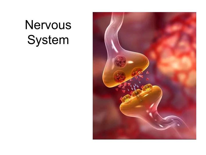
secrete neurotransmitters
Structural Classification
Electrophysiology of neurons
- Key issues
- How does neuron generate
an electrical signal?
- How does a neuron transmit
that signal to the next cell?
Cell Membrane Structure
- phospholipid bilayer
- embedded proteins
Channel Proteins
- nongated
- chemically gated
- neurotransmitter
- voltage gated
Resting membrane potential
- 70mV
- cytosol compared to extracellular fluid
- Negative inside of
cell relative to
- utside
- Anions inside cell:
proteins, nucleic acids, phosphates
- Cations: excess
Na+ outside cell; excess K+ inside cell
Resting membrane potential
- K+ diffuses out
- pulled back
in due to electrical force
- Na+ diffuses
slowly in
- Na+ - K+ pump
counteracts diffusion
Sodium-Potassium Pump
- 3 Na+ pumped out
- 2 K+ pumped in
- Requires ATP
- Na+ and K+
constantly leak back through membrane by diffusion
- Resting
membrane potential =
- 70mV
Neuron stimulation
- Begins at dendrites
- Spreads through the soma
- Travels down the axon
- Ends at the synaptic knobs
Neuron excitation
- signal = change in membrane potential
- alter ion concentration
- alter membrane permeability to ions
- 2 types of signals
- local (graded) potentials
§ incoming, short distance
- action potentials
§ axon signals, long distance
Local (graded) potential
- Stimulation of dendrite by chemicals, light,
heat or mechanical distortion
- Stimulation causes Na+ gates to open
- Na+ rushes into the cell
- Depolarization – shifting membrane potential
Local (graded) potential
- Inside: K+ move away from depolarized area
- Outside: Na+ move toward depolarized area
- Cl- ions take their places
- Depolarization moves away from stimulus area
Characteristics of local potentials
- Vary in magnitude: stronger stimulus opens
more Na+ gates resulting in higher potential
- Decremental: K+ flows out of cell rapidly
after stimulation
- prevents local potential from having long-
distance effects
Characteristics of local potentials
- Reversible – if stimulation stops, resting
membrane potential is quickly restored
Action Potentials (aka nerve impulse)
- Can occur in neurons and skeletal muscle
- Only occurs if excitatory local potential is
strong enough when it arrives at the trigger zone
Action Potentials (aka nerve impulse)
- 3 phases
- depolarization
- repolarization
- hyperpolarization
Action Potential
- Depolarization
- Na+ gates
- pen
- Depolarization
causes more Na+ gates to
- pen (positive
feedback)
- At 0mV, Na+
gates begin closing
Action Potential
- Voltage peaks
between 0-50mV
- Membrane is
now positive
- n the inside
(reverse of resting membrane potential)
Action Potential
- K+ gates have
also been
- pening but
more slowly
- At voltage
peak, K+ gates are fully open
Action Potential
- Repolarization
- K+ exit cell due
to diffusion
- K+ exit cell due
to repulsion by positive charge
- f cytoplasm
- Exiting of K+
brings voltage back down
Action Potential
- Hyperpolarization
- K+ gates stay
- pen longer than
Na+ gates
- Results in drop of
membrane potential below resting state
Action Potential
- Restoration of
resting membrane potential
- Diffusion of ions
through membrane
- Sodium-potassium
pump
Action Potential
Characteristics of action potentials
- Threshold point initiates firing
- depolarization by 15-20mV
- All-or-none law
- if neuron fires, it does so at its maximum
voltage
- Nondecremental
- all action potentials throughout neuron are
same strength
- Irreversible
- action potential cannot be stopped once it
starts
Refractory period
- Period immediately following action potential
- Cannot stimulate that region of the
membrane again
- Lasts until hyperpolarization ends (until K+
channels reclose and Na+ channels recover)
Conduction in unmyelinated fiber
- Depolarization in one part of the
membrane triggers Na+ to open in the adjacent areas of the membrane
- Conduction rate = 2 m/s
- Action potentials are produced
sequentially in adjacent membrane
- Refractory period prevents backflow of
conduction
Myelin
- Insulates
- Mostly lipid (as
cell membrane)
- Oligodendrocyte
- r Schwann cell
- Speeds
conduction of nerve signal
Conduction in myelinated fibers
- 30x faster than unmyelinated
- Myelin insulates membrane from
extracellular fluid
- Ions cannot flow in or out of cell in
myelinated regions
- Ions can flow at nodes of Ranvier
Conduction in myelinated fibers
- Na+ enters at node and diffuses in axon
under myelin sheath
- This signal decreases as it moves down the
axon
- At next node of Ranvier, signal is just strong
enough to generate next action potential
Saltatory conduction
- Internodes
- Diffusion is fast but decremental
- Nodes of Ranvier
- Conduction is slow but nondecremental
Synaptic connections
- Pre-synaptic neuron
- Synaptic cleft
- Neurotransmitter
- Post-synaptic neuron
One neuron can have as many as 100,000 synapses!
Synaptic transmission
- Nerve signal
arrives at synaptic knob
- Ca++ gates open
- Ca++ enters knob
and triggers synaptic vesicles to release neurotransmitter
300 vesicles could be released!
Synaptic transmission
- Neurotransmitter
diffuses across synaptic cleft
- neurotransmitter
binds to gates on post-synaptic neuron
Excitatory Synapse
- Gates open to let
Na+ in and K+ out
- Post-synaptic
membrane depolarizes
- If strong enough,
triggers post- synaptic neuron to fire
Inhibitory Synapse
- Gates open to let
Cl- in and/or K+ out
- Post-synaptic
membrane hyperpolarizes
- Decreased
likelihood of post- synaptic neuron firing
Cessation of the signal
- Neurotransmitter only binds to a receptor for
1msec, then dissociates from it
- Neurotransmitters diffuse away from the
synaptic cleft and get reabsorbed (by astrocytes)
- Synaptic knobs reuptake
neurotransmitters
- Enzymes in the synaptic cleft break down
neurotransmitters
for a more or less complete list see: http://wiki.answers.com/Q/List_all_the_essential_neurotransmitters
Sensory Receptors
Classification by location
- mechanoreceptors (touch)
- photoreceptors (light)
- thermoreceptors (heat)
- chemoreceptors (chemical)
- nociceptors (pain)
Sensory Receptors
Classification by location
- exteroceptors
- stimulus outside body
- interoceptors
- stimulus inside body
- proprioceptors
- interoceptors for body movement/stretch
§ skeletal muscle § tendons § ligaments § connective tissue over bones and muscles
Integration
- 3 basic levels
- receptor level
- sensory reception
- transmission to CNS
- circuit level
- processing in
ascending pathways
- perceptual level
- processing in the
cortex
Reflex arcs
visceral (note that integration may be within wall of GI tract somatic note that both visceral and somatic pain travel the same afferent pathway