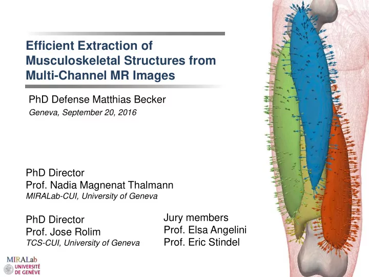

Efficient Extraction of Musculoskeletal Structures from Multi-Channel MR Images PhD Defense Matthias Becker Geneva, September 20, 2016 PhD Director Prof. Nadia Magnenat Thalmann MIRALab-CUI, University of Geneva Jury members PhD Director Prof. Elsa Angelini Prof. Jose Rolim Prof. Eric Stindel TCS-CUI, University of Geneva
Outline Introduction Related work Contributions in four areas 1. Image acquisition 2. Image processing 3. Model initialisation 4. Image segmentation Conclusions Conclusions, limitations and future work 2 PhD Defense Matthias Becker
Research problem • Introduction • Related Work • Contributions • Conclusions Musculoskeletal diseases 34000000 29000000 morphology of anatomical 24000000 # of scans structures explain some of the 19000000 14000000 origins 9000000 year 1995 1996 1997 1998 1999 2000 2001 2002 2003 2004 2005 2006 2007 2008 2009 2010 2011 MRI MRI scans (USA) based on OECD data Highlights multiple structures High contrast in soft tissue Number of MR scans increases Generates big amounts of data Multi-channel acquisition 3 PhD Defense Matthias Becker
Research problem • Introduction • Related Work • Contributions • Conclusions Development of an imaging protocol Tissue labelling in image data Exploitation of multi-channel image data during the segmentation 4 PhD Defense Matthias Becker
Outline Introduction Related work Contributions in four areas 1. Image acquisition 2. Image processing 3. Model initialisation 4. Image segmentation Conclusions Conclusions, limitations and future work 5 PhD Defense Matthias Becker
Muscle segmentation • Introduction • Related Work • Contributions • Conclusions Example based muscle segmentation Method for a semi-automatic muscle segmentation User manually outlines the contour • in pivot slices Interpolation and optimization [JDR+14] E. Jolivet, E. Dion, P. Rouch, G. Dubois, R. Charrier, C. Payan, and W. Skalli , “Skeletal muscle segmentation from MRI dataset using a model- based approach,” Comput. Methods Biomech. Biomed. Eng. Imaging Vis. , no. April 2014, pp. 1 – 8, Apr. 2014. 6 PhD Defense Matthias Becker
Muscle segmentation • Introduction • Related Work • Contributions • Conclusions Complex task, muscles hard to distinguish Limit to compartments [BACP12] Previous work shows [TNL+14] problems in accuracy [BACP12] and performance [G07] [BACP12] Baudin, P.Y., Azzabou, N., Carlier, P.G., Paragios, N.: Prior knowledge, random walks and human skeletal muscle segmentation. MICCAI. 15, 569 – 76 (2012). [G07] Gilles, B.: Anatomical and Kinematical Modelling of the Musculoskeletal System from MRI, [G07] Thesis, (2007). [TNL+14] M. S. Thomas, D. Newman, O. D. Leinhard, B. Kasmai, R. Greenwood, P. N. Malcolm, A. Karlsson, J. Rosander, M. Borga , and A. P. Toms, “Test -retest reliability of automated whole body and compartmental muscle volume measurements on a wide bore 3T MR system.,” Eur. Radiol. , May 2014. 7 PhD Defense Matthias Becker
Muscle segmentation • Introduction • Related Work • Contributions • Conclusions Statistical Shape Models Andrews et al. : SSM approach for muscle segmentation in the thigh Alignment and PCA from 39 data sets Segmentation uses energy minimization using image features (intensities) and derived features (gradients, curvature). Avg. DSC of 0.92 [AHY+11] S. Andrews, G. Hamarneh, A. Yazdanpanah, B. Haj Ghanbari , and W. D. Reid, “Probabilistic multi -shape segmentation of knee extensor and flexor muscles.,” Med. Image Comput. Comput. Assist. Interv. , vol. 14, no. Pt 3, pp. 651 – 8, Jan. 2011. 8 PhD Defense Matthias Becker
Multi-channel segmentation • Introduction • Contributions • Conclusions • Related Work Multi-channel applications [CSV00] Edge-less level sets [KJC13] on RGB images [F11] - Left ventricle in CT and PET [GCM+11] [F11] Fechter, T. Deformation Based Manual Segmentation in Three and Four Dimensions ., 2011 [CSV00] Chan, T. F., Sandberg, B. Y., & Vese, L. a. (2000). Active Contours without Edges for Vector-Valued Images. Journal of Visual Communication and Image Representation , 11 (2), 130 – 141. doi:10.1006/jvci.1999.0442 [KJC13] I. Kopriva, A. Jukić, and X. Chen, “Sparseness constrained nonnegative matrix factorization for unsupervised 3D segmentation of multichannel images: demonstration on multispectral magnetic resonance image of the brain,” vol. 8669,Mar. 20 13. [GCM+11] E. Geremia, O. Clatz, B. H. Menze, E. Konukoglu, A. Criminisi, and N. Ayache , “Spatial decision forests for MS lesion segmentation in multi- channel magnetic resonance images.,” Neuroimage , vol. 57, no. 2, pp. 378 – 90, Jul. 2011. 9 PhD Defense Matthias Becker
Related work • Introduction • Related Work • Contributions • Conclusions Model based approaches have high potential in musculoskeletal segmentation Comprehensive approach, including anatomical knowledge [AHY+11] Multi-channel provides additional image information Results in higher segmentation quality [BFS+07] Our proposal: Multi-channel + Deformable Models Need for higher efficiency: Larger amount of data requires more processing [AHY+11] S. Andrews, G. Hamarneh, A. Yazdanpanah, B. Haj Ghanbari , and W. D. Reid, “Probabilistic multi -shape segmentation of knee extensor and flexor muscles.,” Med. Image Comput. Comput. Assist. Interv. , vol. 14, no. Pt 3, pp. 651 – 8, Jan. 2011. [BFS+07] P. Bourgeat, J. Fripp, P. Stanwell, S. Ramadan, and S. Ourselin , “MR image segmentation of the knee bone using phase information,” Med. Image Anal. , vol. 11, no. 4, pp. 325 – 335, 2007. 10 PhD Defense Matthias Becker
Outline Introduction Related work Contributions in four areas 1. Image acquisition 2. Image processing 3. Model initialisation 4. Image segmentation Conclusions Conclusions, limitations and future work 11 PhD Defense Matthias Becker
Contributions • Introduction • Related Work • Contributions • Conclusions 2. Image processing 3. Model initialisation 4. Segmentation 1. Image acquisition 12 PhD Defense Matthias Becker
1. Image acquisition • Introduction • Related Work • Contributions • Conclusions MRI Acquisition [GDT+14] Surface coil • Higher resolution, shorter duration, better SNR Only covers 30cm coil Subject must not move scanner wooden board scanner table [GDT+14] D. García, B. M. A. Delattre , S. Trombella, S. Lynch, M. Becker, H. F. Choi, and O. Ratib, “Open framework for management and processing of multi- modality and multidimensional imaging data for analysis and modeling muscular function,” Proc. SPIE 9036 , Medical Imaging 2014: Image-Guided Procedures, Robotic Interventions, and Modeling, 90361W (March 12, 2014). 13 PhD Defense Matthias Becker
1. Image acquisition • Introduction • Related Work • Contributions • Conclusions [GDT+14] D. García, B. M. A. Delattre , S. Trombella, S. Lynch, M. Becker, H. F. Choi, and O. Ratib, “Open framework for management and processing of multi- modality and multidimensional imaging data for analysis and modeling muscular function,” Proc. SPIE 9036 , Medical Imaging 2014: Image-Guided Procedures, Robotic Interventions, and Modeling, 90361W (March 12, 2014). 14 PhD Defense Matthias Becker
1. Image acquisition • Introduction • Related Work • Contributions • Conclusions Multi-channel acquisition: mDixon and T1/TSE mDixon T1/TSE opposed- in- phase fat phase water magnitude 29 to 52 minutes (3-5 stacks) Static scan Knee scan Stack Stack Stack Move setup: 4:00 Move setup: 4:00 Move setup: 5:00 3D DP Vista: 3:16 T1 TSE cor: 2:00 Reference: 0:31 Reference: 0:31 Reference: 0:31 Reference: 0:31 Survey: 0:10 T1 TSE: 4:51 Survey: 0:10 T1 TSE: 4:51 Survey: 0:10 T1 TSE: 4:51 Survey: 0:50 Dixon: 1:42 Dixon: 1:42 Dixon: 1:42 … t 15 PhD Defense Matthias Becker
1. Image acquisition • Introduction • Related Work • Contributions • Conclusions Study subjects Subject 1 Subject 2 Subject 3 Subject 4 Subject 5 Age 26 24 29 22 32 Height 175 172 197 176 173 Weight 85 65 84 68 58 Activity None Running Running Soccer Soccer 16 PhD Defense Matthias Becker
1. Image acquisition • Introduction • Related Work • Contributions • Conclusions Air labelling Addition Can be used to crop files Threshold Connected Uncropped Cropped Reduction component S1 1877 MiB 701 MiB 62 % S2 1973 MiB 571 MiB 71 % 4x Dilation S3 2133 MiB 728 MiB 66 % 4x Erosion S4 1998 MiB 684 MiB 66 % Inversion S5 2418 MiB 658 MiB 72 % Connected component 17 PhD Defense Matthias Becker
2. Image processing • Introduction • Related Work • Contributions • Conclusions 2. Image processing 3. Model initialisation 1. Image acquisition 4. Segmentation 18 PhD Defense Matthias Becker
Recommend
More recommend