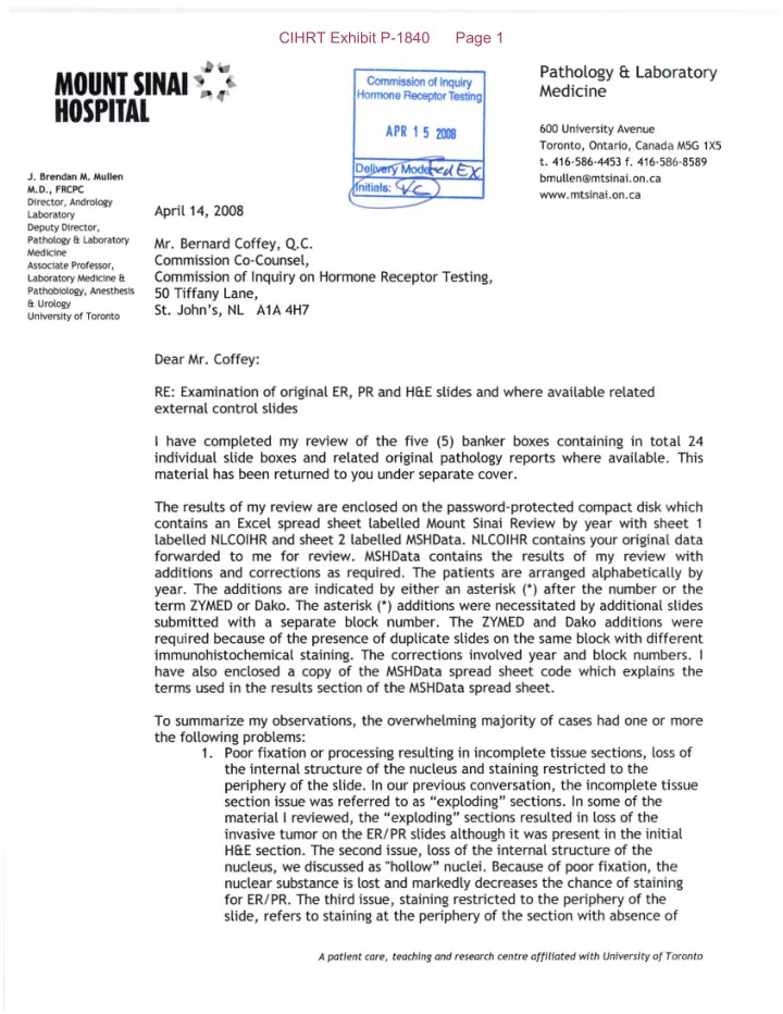

CIHRT Exhibit P-1840 Page 1 Pathology ft Laboratory MOUNT SINAI Commission 01 Inquiry Medicine Hormone Receptor Testing HOSPITAL 600 University Avenue APR 1 5 ZOOS Toronto, Ontario, Canada M5G 1X5 t. 416·586-4453 f. 416-586-8589 J. Brendan M. Mullen bmullen@rntsinai.on.ca M.D., FRCPC www.mtsinai.on.ca Director, Andrology April 14, 2008 Laboratory Deputy Director, Pathology &. laboratory Mr. Bernard Coffey, Q.c. Medicine Commission Co-Counsel, Associate Professor, Commission of Inquiry on Hormone Receptor Testing, Laboratory Medicine &. Pathobiology, Anesthesis 50 Tiffany Lane, &. Urology St. John's, NL AlA 4H7 University of Toronto Dear Mr. Coffey: RE: Examination of original ER, PR and H&E slides and where available related external control slides I have completed my review of the five (5) banker boxes containing in total 24 individual slide boxes and related original pathology reports where available. This material has been returned to you under separate cover. The results of my review are enclosed on the password-protected compact disk which contains an Excel spread sheet labelled Mount Sinai Review by year with sheet 1 labelled NLCOIHR and sheet 2 labelled MSHData. NLCOIHR contains your original data forwarded to me for review. MSHData contains the results of my review with additions and corrections as required. The patients are arranged alphabetically by year. The additions are indicated by either an asterisk (') after the number or the term ZYMED or Dako. The asterisk (') additions were necessitated by additional slides submitted with a separate block number. The ZYMED and Dako additions were required because of the presence of duplicate slides on the same block with different immunohistochemical staining. The corrections involved year and block numbers. I have also enclosed a copy of the MSHData spread sheet code which explains the terms used in the results section of the MSHData spread sheet. To summarize my observations, the overwhelming majority of cases had one or more the following problems: 1. Poor fixation or processing resulting in incomplete tissue sections, loss of the internal structure of the nucleus and staining restricted to the periphery of the slide. In our previous conversation, the incomplete tissue section issue was referred to as "exploding" sections. In some of the material I reviewed, the "exploding" sections resulted in loss of the invasive tumor on the ER/PR slides although it was present in the initial H&E section. The second issue, loss of the internal structure of the nucleus, we discussed as "hollow" nuclei. Because of poor fixation, the nuclear substance is lost and markedly decreases the chance of staining for ER/PR. The third issue, staining restricted to the periphery of the slide, refers to staining at the periphery of the section with absence of A patient care, teaching and research centre affiliated with University of Toronto
CIHRT Exhibit P-1840 Page 2 staining centrally. It is difficult to interpret the results of these cases as the peripheral staining results may not reflect the results of the entire tumour. 2. Absence of the internal controls. Many of the cases had no normal duct epithelium to use as an internal control on the initial Hf1E section. Additionally, in many cases where the original Hf1E section had normal duct epithelium, it was not present on the ER/PR slides as a result of the "exploding" section issue. 3. Negative internal controls. In many cases, the internal control either did not stain or stained very weakly. Also, with the exception of a small minority of cases, the ER internal control was significantly weaker than the PR internal control. 4. Stain deposit obscuring morphology. In many cases excess stain was present either on the surface or beneath the section. Both artifacts preclude assessment of the ER/PR staining in areas affected. 5. External controls. The external controls were inconsistent both between slides and within slides. In some cases, the positive cells were barely stained. In occasional cases from 2005, the controls stained both the nucleus and the cytoplasm reflecting inadequate or incorrect validation. 6. Discrepancy between internal and external controls. In only one or two of the 539 cases I reviewed was the staining in the internal control as strong as the corresponding external control. There were very few cases in which there was a significant difference in my observation compared to that recorded on the original report. Some of my observations were higher than those recorded and some lower. Please contact me at 416-586-4553 for the password. If you have any questions regarding the MSHData spread sheet, the MSHData spread sheet code or my observations, please do not hesitate to contact me. Yours truly, J. Brendan M. Mullen, MD JBMM/mtm Pase 2 af 2
CIHRT Exhibit P-1840 Page 3 MSHData Spread Sheet Code: MSHReview Tumour type D - ductal DL - ductal with lobular features L -lobular DCISIM - ductal carcinoma in situ with microinvasion «lmm) DC - ductal with colloid features C - colloid MCa - metastatic carcinoma EPAP - encysted papillary in situ carcinoma PAP - papillary carcinoma NT - no tumour L YG - Iymphangitic carcinoma No H&E - no routine slide to assess tumour and tumour not present on ERiPR slides ER - % cells positive PR - % cells positive IC - internal controls with P-present but not stained, PS-present and stained, PSW-present and stained weakly, A-absent. The value refers to the ER internal control. [fthe ER and internal control was not present within the value refers to be PR internal control. The comment in the latter case will state ER IC NEG (estrogen receptor internal control negative). F/P - fixation and processing with A-adequate and P-poor EC - external controls slides when available with P - positive, ?P - questionable positive. If both ER and PR external control slides were present they were reported as PIP, if only the ER control slide was present it was reported as P, if only the PR control slide was present it was reported as IP and if two sets of control slides were present they were reported as P/PIPIP. No external control was negative. NL Original ER & PR - The results present on the accompanying report when provided. If a numerical value was not reported then the qualitative result was entered (N - negative, MP - moderately positive, WP - weakly positive, OP - occasionally positive and RARE - rare positive). One PR result measured by biochemical means was reported as E - equivocal. Comment - When a surgical pathology report was provided which included the results of the MSH retrospective review, I compared the NL results to the MSH results. The results were categorized as CONCORDANT if the two results were in the same ER group and DISCORDANT if they were in different ER groups [negative «1%), low positive (1- 10%) and positive (>10%)].
Recommend
More recommend