
SLIDE 1
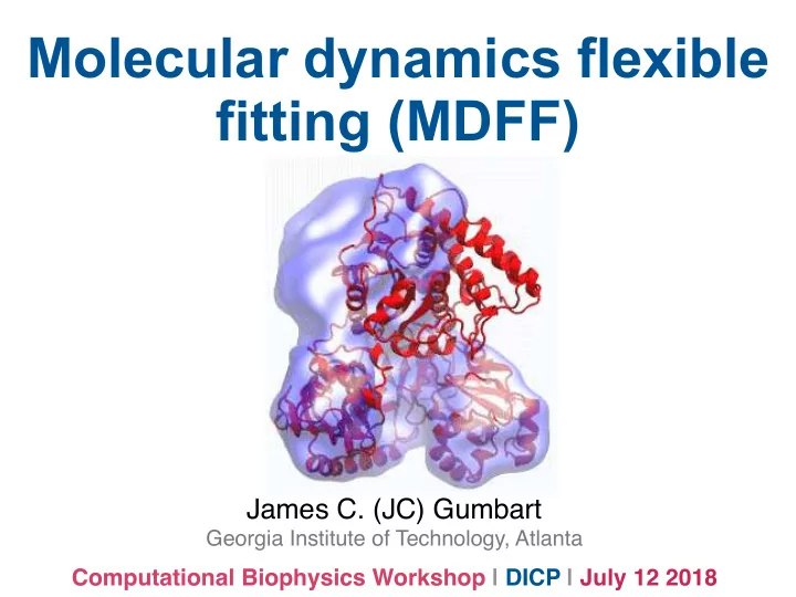
Molecular dynamics flexible fitting (MDFF)
James C. (JC) Gumbart
Georgia Institute of Technology, Atlanta
Computational Biophysics Workshop | DICP | July 12 2018
Molecular dynamics flexible fitting (MDFF) James C. (JC) Gumbart - - PowerPoint PPT Presentation
Molecular dynamics flexible fitting (MDFF) James C. (JC) Gumbart Georgia Institute of Technology, Atlanta Computational Biophysics Workshop | DICP | July 12 2018 Structural biology continuum 1 cm 1 m 1 mm 1 nm 1 Light microscopy
Georgia Institute of Technology, Atlanta
Computational Biophysics Workshop | DICP | July 12 2018
1 cm 1 mm 1 μm 1 nm 1 Å Light microscopy Electron microscopy X-ray, NMR Modeling and molecular dynamics
resolution of the resulting image is limited* by the wavelength of light used
d = λ 2(n sin θ)
Abbe diffraction limit
*except in super-resolution imaging
The Nobel Prize in Chemistry 2017 was awarded … "for developing cryo-electron microscopy for the high-resolution structure determination of biomolecules in solution".
2D images are aligned and sorted computationally into classes representing homogeneous particles and perspectives
h"p://people.csail.mit.edu/gdp/cryoem.html
classes are then averaged and back-projected to produce 3D density map
h"p://people.csail.mit.edu/gdp/cryoem.html
back projection is iterative - need the model for projection matching with class averages cryo-EM map of the proteosome (iteration 1) final map maps can have resolutions ranging from near-atomic (<5 Å) to 2-3 nm
Split dataset in half, calculate two independent reconstructions Align two structures, flip into reciprocal space (i.e., 3D FT), and calculate correlation co- efficients between bands of spatial frequency
Fourier shell correlation:
FSC between two halves of the data set
i
using Molecular Dynamics Flexible Fitting (MDFF)
Two terms are added to the MD potential A mass-weighted force is then applied to each atom An external potential derived from the EM map is defined on a grid as
Flexible fitting of atomic structures into electron microscopy maps using molecular dynamics. L G. Trabuco*, E Villa*, K Mitra, J Frank, K Schulten. Structure, 16, 673-683, 2008.
Arrows (representing forces) point to regions of higher density (lower energy)
Flexible fitting of atomic structures into electron microscopy maps using molecular dynamics. L G. Trabuco*, E Villa*, K Mitra, J Frank, K Schulten. Structure, 16, 673-683, 2008.
USS = X
restraints
kµ(µ − µ0)2
Harmonic restraints are applied to preserve secondary structure of proteins and nucleic acids, avoiding “overfitting” For proteins, ɸ and ѱ dihedral angles
are restrained. Hydrogen-bond restraints are also an option.
Flexible fitting of atomic structures into electron microscopy maps using molecular dynamics. L G. Trabuco*, E Villa*, K Mitra, J Frank, K Schulten. Structure, 16, 673-683, 2008.
USS = X
restraints
kµ(µ − µ0)2
Harmonic restraints are applied to preserve secondary structure of proteins and nucleic acids, avoiding “overfitting” For nucleic acids, distance and dihedral restraints are applied to a selected set of base pairs.
Flexible fitting of atomic structures into electron microscopy maps using molecular dynamics. L G. Trabuco*, E Villa*, K Mitra, J Frank, K Schulten. Structure, 16, 673-683, 2008.
Simulated maps used as targets for proteins with crystal structures in two conformations
Ways to evaluate the quality and convergence of the fit are to track RMSD and cross-correlation coefficient (CCC) Fluctuations about the best fit (“ensemble” of fitted structures)
GroEL-GroES 7-fold Nitrilase helical Mm-cpn 16-fold
parameters, pitch (rotation about central axis) and rise
map to define transformation matrices Ui
← →
U = cos θ − sin θ sin θ cos θ 1 ∆z 1
NAMD can also guess the parameters
red - fit without symmetry blue - fit with symmetry
Symmetry-restrained flexible fitting for symmetric EM
xMDFF: Molecular dynamics flexible fitting of low-resolution X-Ray structures.R McGreevy*, A Singharoy*, Q Li, J Zhang, D Xu, E Perozo, K Schulten. Acta Crystallographica D 70 2344-2355, 2014.
xMDFF: Molecular dynamics flexible fitting of low-resolution X-Ray structures.R McGreevy*, A Singharoy*, Q Li, J Zhang, D Xu, E Perozo, K Schulten. Acta Crystallographica D 70 2344-2355, 2014.
Sec61 in each?
20055 20076
4Menetret, J.F. et al. (2005) J. Mol. Biol. 348:445-457. 5Mitra, K. et al. (2005) Nature 438:318-324. 6Menetret, J.F. et al. (2007) Mol. Cell 28:1083-1092.
20054 20012 19971
1Beckmann, R. et al. (1997) Science 278:2123-2126. 2Beckmann, R. et al. (2001) Cell 107:361-372. 3Morgan, D.G. et al. (2002) J. Mol. Biol. 324:871-886.
20023
SecY (95% of all MPs in eukaryotes)
through SecY’s lateral gate
lateral gate
ribosome membrane exit tunnel closed
Top Side SecY SecY
plug
Low-resolution Data High-resolution Structure Close-up of Nascent Protein in SecY
*collaboration with cryo-EM lab of Roland Beckmann
Unbiased simulation reveals spontaneous attraction of lipids to H59 of ribosome’s 23S...
permitting easier access of the signal anchor to the bilayer
...which explains ambiguous density observed in map!
is the signal anchor where we think it is? New construct made in Rapoport lab (Harvard U.) has disulfide cross-link between plug and nascent chain link confirmed biochemically
C.W. Akey. (2014) Nature. 506:102-106.
resolution 10.1 Å side view PCC SSU LSU
maps by Chris Akey, Boston U.
resolution 9.5 Å Empty complex also visualized
C.W. Akey. (2014) Nature. 506:102-106.
C.W. Akey. (2014) Nature. 506:102-106.
C.W. Akey. (2014) Nature. 506:102-106.
Structures roughly similar, validating placement of signal anchor in first one Frauenfeld et al. 2011 Park et al. 2014 Largest differences are in improved modeling of E. coli- specific elements, e.g., two-helix plug (h2a+h2z) and linker h7b between TMs 7 and 8
h7b h2a + h2z
Nascent chain density is
a V-shaped cleft on top of SecY force from peptide synthesis insufficient to drive translocation
Translocation through the channel may require a pulling force from the other side (e.g., SecDF) Membrane insertion has been demonstrated to exert a force on the nascent chain1
force? force?
1 N. Ismail...G. von Heijne. A biphasic pulling
force acts on transmembrane helices during translocon-mediated membrane insertion. (2012) Nat. Struct. Mol. Bio. 10:1018-1022.
cryo-EM map (25-Å resolution) showed a bent structure (grey outline) in agreement with previous simulations Molecular dynamics flexible fitting of LH1 to that map in the presence of a membrane induced membrane curvature curvature was maintained even after fitting
Jen Hsin, James Gumbart, Leonardo G. Trabuco, Elizabeth Villa, Pu Qian, C. Neil Hunter, and Klaus Schulten. Protein-induced membrane curvature investigated through molecular dynamics flexible fitting. Biophys. J., 97:321-329, 2009.
cryo tomogram of receptor array MDFF fit of atomic structure to averaged map (>30 Å resolution)
Briegel, Ames, Gumbart et al. The mobility of two kinase domains in the Escherichia coli chemoreceptor array varies with signalling state (2013) Mol Microbio. 89:831-841.
The KS group maintains a detailed website with details, examples, and documentation Over 100 published applications in last decade - look for ones similar to the problem or system you are interested in!