MOL2NET, 2017 , 3, doi:10.3390/mol2net-03-xxxx 2 Introduction - - PDF document

mol2net 2017 3 doi 10 3390 mol2net 03 xxxx 2 introduction
SMART_READER_LITE
LIVE PREVIEW

MOL2NET, 2017 , 3, doi:10.3390/mol2net-03-xxxx 2 Introduction - - PDF document

MOL2NET, 2017 , 3, doi:10.3390/mol2net-03-xxxx 1 MOL2NET, International Conference Series on Multidisciplinary Sciences MDPI http://sciforum.net/conference/mol2net-03 Employment of hyphenated approach for metabolomics fingerprinting of


slide-1
SLIDE 1

MOL2NET, 2017, 3, doi:10.3390/mol2net-03-xxxx 1

MDPI

MOL2NET, International Conference Series on Multidisciplinary Sciences http://sciforum.net/conference/mol2net-03

Employment of hyphenated approach for metabolomics fingerprinting of phenolics from Torilis leptophylla roots

Noshin Nasreen (noshinnasreen@yahoo.com) a, Nabil Semmar (nabilsemmar@yahoo.fr) b, Muhammad Farman(farman@qau.edu.pk) a, Naseem Saud Ahmad (saudahmad@uhs.edu.pk)c

a Department of Chemistry, Quaid-i-AzamUniversity, Islamabad-45320,Pakistan bDepartment of Bioinformatics, Biomathematics & Biostatistics, University of Tunis El Manar, Tunis,

Tunisia

cDepartment of Pharmacology University of Health Sciences, Lahore

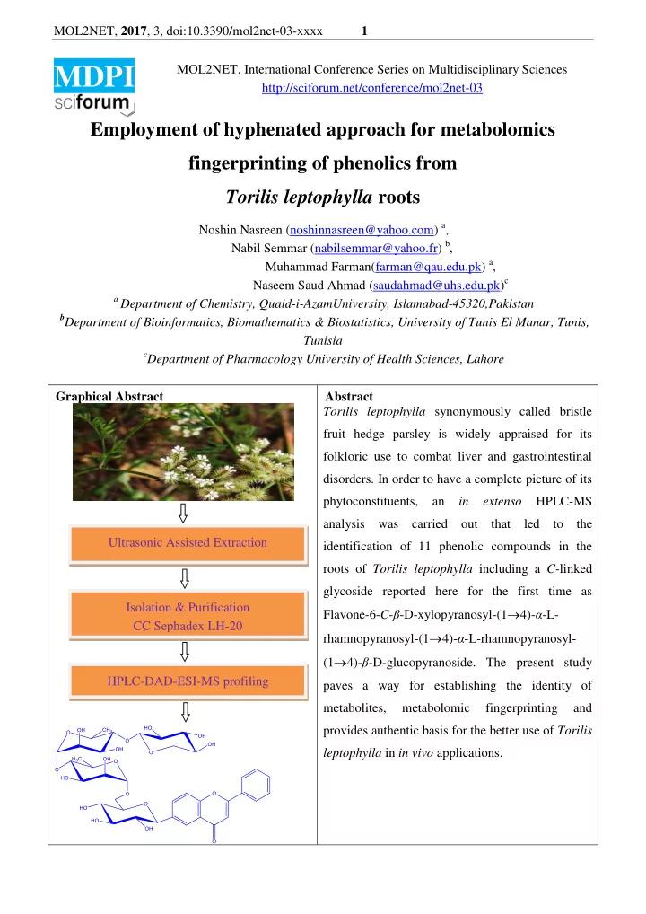
Graphical Abstract Abstract Torilis leptophylla synonymously called bristle fruit hedge parsley is widely appraised for its folkloric use to combat liver and gastrointestinal

  • disorders. In order to have a complete picture of its

phytoconstituents, an in extenso HPLC-MS analysis was carried out that led to the identification of 11 phenolic compounds in the roots of Torilis leptophylla including a C-linked glycoside reported here for the first time as Flavone-6-C-β-D-xylopyranosyl-(14)-α-L- rhamnopyranosyl-(14)-α-L-rhamnopyranosyl- (14)-β-D-glucopyranoside. The present study paves a way for establishing the identity of metabolites, metabolomic fingerprinting and provides authentic basis for the better use of Torilis leptophylla in in vivo applications. Ultrasonic Assisted Extraction Isolation & Purification CC Sephadex LH-20 HPLC-DAD-ESI-MS profiling

slide-2
SLIDE 2

MOL2NET, 2017, 3, doi:10.3390/mol2net-03-xxxx 2 Introduction Torilis species (Apiaceae), popular worldwide as hedge parsleys, comprises 11-13 species of aroma containing plants possessing diverse phytoconstituents and pronounced biological activities. Only three species of genus Torilis has been reported from various domains of Pakistan [1]. Torilis leptophylla is an annual herb that is traditionally a well known medication to relieve hepatic, gastric and intestinal disorders. Due of its potential to act as a disinfectant, the plant is highly efficacious against some pathogens [2]. Furthermore Torilis leptophylla leaves are taken as vegetable while stem and branches serve as fodder [3]. The comprehensive profile of Torilis leptophylla roots has not been documented earlier. In

  • rder to discern the full medicinal prospects of Torilis leptophylla roots, it is required to have a

complete picture of its phytoconstituents therefore, the present study aimed at characterization, isolation and metabolomic fingerprinting of phenolics using HPLC-DAD-ESI-MS analysis. Materials and Methods Extraction Torilis leptophylla roots extracts were prepared by infusion technique. Plant material (100 grams) was added to 300 mL of methanol: water (70: 30). Contents were irradiated at 35 KHz frequency and 220 volts by using Elma Ultrasonic LC-30H instrument for 15 minutes and stored at room temperature. After 24 hours infusions were filtered and the whole process was repeated thrice. Combined supernatants were evaporated to dryness in vacuo using Heidolph 4000-efficient rotary

  • evaporator. As a consequence, crude extract having syrupy consistency was obtained.

Acid Hydrolysis 5 gm Torilis leptophylla roots were added to 2M HCl and methanol in (1:1) and contents were refluxed on a boiling water bath for 3 hours. Extract was cooled, filtered and filtrate was extracted thrice, each time with 25 mL of ethyl acetate to separate the pool of aglycones and sugars. Aqueous and ethyl acetate layers were separated and evaporated to dryness on a rotary evaporator. Aglycones (Agly) remained in ethyl acetate layer whereas sugars (Hyd) moved to the aqueous layer. Spots of standard sugars and Hyd were applied on the TLC plate, developed, sprayed and heated in an

  • ven at 105 0C for 5-10 min. until the brownish spots appeared. The sugars present in the sample were

identified by comparison of their Rf values with those of the standard sugars [4]. The sugars were identified by using precoated silica gel 60F254 (Merck) as a stationary phase. Column chromatography Column chromatography was carried out by using Sephadex LH-20, a dextran gel that swells in water resulting in shrinkage of pore volume. Exploring the size exclusion chromatographic technique, slurry of sephadex made in MeOH : H2O (70:30) was introduced into column having dimensions 17  1.5 cm, plugged with glass wool. 1.5 mL of herbal extract was introduced on the top of sephadex.

slide-3
SLIDE 3

MOL2NET, 2017, 3, doi:10.3390/mol2net-03-xxxx 3 Elution was carried out by the same solvent and monitored by UV. Separation took place on the basis

  • f molecular sizes. Larger molecules being eluted first followed by smaller sized molecules. Different

fractions were collected which were later subjected to HPLC-DAD-ESI-MS analysis. HPLC-DAD-ESI-MS analysis Parameters The flavonoids composition of Torilisleptophylla was determined by injecting 5 µL of extract in Agilent Technology 1200 LC instrument’s stainless steel column (4.6 X 150mm) packed with 5 µmthickness of Agilent eclipse extra dense bonding (XDB) reverse phase C18 silica. Elution was carried out by using binary solvent system mobile phase (deionized water labeled as solvent A and HPLC grade acetonitrile labeled as solvent B). 0.1% formic acid was added in both the solvents. Sixty minutes gradient elution program was followed as 10% of acetonitrile at 0 minutes, 10% of acetonitrile at 15 minutes, 40% of acetonitrile at 40 minutes, 80% of acetonitrile at 50 minutes and 10% of acetonitrile at 60 minutes. Flow of mobile phase was maintained at a rate of 0.5 mL/min. Diode array detector was adjusted to detect three different wavelengths (254 nm, 320 nm and 370 nm). On the

  • ther hand mass spectrometer was equipped with electrospray ionization mode (ESI), Ion trap analyzer

and photomultiplier tube as detector. Spectra were recorded in negative ionization mode between mass range of 50-2200 a.m.u. Samples preparation Clear concentrated hydroalcoholic extracts were subjected to HPLC-DAD-ESI-MS analysis to explore the phenolic compounds. Results and Discussion HPLC-DAD-ESI-MS Analysis HPLC Chromatogram of the isolated fraction of hydro-alcoholic extract of TL is shown in fig.1. This pure fraction was obtained from column chromatography using sephadex as a stationary phase and MeOH : H2O (70:30) as mobile phase. Fig.1 HPLC chromatogram of isolated fraction TL-F6 Compound eluted at retention time of 32.4 min when the composition of mobile phase was 28% acetonitrile in deionized water. More proportion of water revealed more hydrophilic nature of the eluted compound. i.e., tetraglycosides. DAD response showed characteristic band pattern of flavones where band II corresponding to ring A appeared at 240 nm. The intensity of band I was higher as compared to band II. Hypsochromic shift of 10 nm from normal value indicated glycosylation on ring

slide-4
SLIDE 4

MOL2NET, 2017, 3, doi:10.3390/mol2net-03-xxxx 4

  • A. Typical value of absorption of band I at 330 nm indicated no substitution on B ring.The shape,

intensities and λmax values of the two bands were closely related to flavone-6-C-tetraglycoside [5]. Spectral data for compound TL-F6 is shown in fig. 2. (a) (b)

  • Fig. 2 Spectral data for compound TL-F6 a)DAD response b) ESI-MS spectrum
  • f compound TL-F6

The ESI-MS spectrum of the compound TL-F6 recorded in the –ve ionization mode showed deprotonated molecular ion at m/z 807. Sequential losses of 132, 146, 146 and 149 mass units probably justified the consecutive attachment of Sugars. The pictorial representation of TLC and PC analyses of sugars and aglycones obtained from acid hydrolysis is shown in fig. 3. Stationary phase= Silica gel 60F254(Merck) impregnated with Sodium dihydrogen phosphate Mobile phase = Acetone :Water (9:1) Spraying reagent =Aniline hydrogen phthalate Stationary phase = Cellulose Mobile phase 1D = BAW (4:1:5) 2D = 15% AcOH

  • Fig. 3 TLC and PC analyses of Hyd and AGLY obtained from acid hydrolysis

The TLC and PC analyses of the hydrolysate provided extra information in probing the structure of glycoside. Nature of sugars was determined from mass losses however the identity was established from co-chromatography of hydrolysate. Loss of pentose, hexose and deoxyhexose sugars were assigned to xylose, glucose and rhamnose respectively.

Rf = 0.41 Rf = 0.60 Rf = 0.70 Rf = 0.80 Rf = 0.92

Rha Xyl Ara Glc Gal Hyd

C-linked glycoside

slide-5
SLIDE 5

MOL2NET, 2017, 3, doi:10.3390/mol2net-03-xxxx 5 The PC analysis provided supplementary information regarding the C-linked nature of

  • glycosides. Loss of 149 mass units and additional 13 mass units with aglycone fragment disclosed the

C-linked nature of aglycone. Linkages between the sugars were determined on the basis of literature cited. In nature [14] linkage of xylose to any other sugar, [14] linkage of rhamnose to glucose and [14] linkage between two rhamnose moieties is known. The structure was determined keeping in view the DAD response, the differences in m/z, sequential losses of sugars and deductions from TLC and PC analyses. Deprotonated molecular ion appeared at m/z 807 [M-H]-and it determined m/z 808 as the molecular mass of the compound. The fragment at m/z 717 indicated 0,2X cleavage of xylose i.e., [M- H-0,2Xxyl]-[6].The peak corresponding to m/z 675 indicated the cleavage of xylose moiety i.e., [M-H- xyl]-. Signal at m/z 529 resulted due to sequential cleavage of xylose and rhamnose from the deprotonated molecular ion i.e.,[M-H-xyl-rham]-. Fragment corresponding to m/z 383 indicated losses

  • f xylose and two rhamnose moieties from the compound i.e., [M-H-xyl-rham-rham]-. Appearance of

peak at m/z 283 suggested the sequential losses of one xylose, two rhamnose and a C-linked glucose moiety from the compound i.e., [M-H-xyl-rham-rham-glc(C-linked)]-. The justification of major fragments formed in ESI-MS further ensured that the compound was Flavone-6-C-β-D-xylopyranosyl- (14)-α-L-rhamnopyranosyl-(14)-α-L-rhamnopyranosyl-(14)-β-D-glucopyranoside. Spectral data for the characterization of compounds in Torilis leptophylla roots by HPLC-DAD-ESI-MS analysis is shown in table 1. Table 1: Spectral data for the characterization of compounds in Torilis leptophylla roots by HPLC- DAD-ESI-MS analysis

Compounds Codes DAD λmaxvalue Pseudo molecular Ion Fragment ions Identified compounds

TLF-6 240,330 [M-H]- 807,717,675,608, 529,383,233 Flavone-6-C-β-D-xylopyranosyl-(14)-α-L- rhamnopyranosyl-(14)-α-L-rhamnopyranosyl- (14)-β-D-glucopyranoside TLF-7 230,325 [M-H] - 693,531,427,369 Flavone-7-O-β-D-glucopyranosyl-(16)- β-D- glucopyranosyl-(14)- β-D-xylopyranoside TLF-8 220, 240sh, 326 [M-2H+ACN]- 887,643,319 Ampelopsin-7-O-α-L-3״-propanoylrhamnopyranosyl- (16)-β-D-3״-caffeylglucopyranoside

slide-6
SLIDE 6

MOL2NET, 2017, 3, doi:10.3390/mol2net-03-xxxx 6

TLF-9 230,320 [M-H]- 563,503,329,269, 239 Apigenin-7-O-β-D-xylopyranosyl-(14)-β-D- glucopyranoside TLF-10 268, 342 [M-H+H2O]- 889,737,685,563, 269 Apigenin-7-O-β-D-glucopyranosyl-(14)-β-D- rhamnopyranosyl-(16)-β-D-glucopyranosyl- (14)-β-D-xylopyranoside TLF-11 240, 340 [M-H] - 563,473,269 Apigenin-4-O-β-D-glucopyranosyl-(14)-β-D- xylopyranoside TLF-12 248, 350 [M-H+H2O]- 595,477,449,269 Apigenin-7-O-rutinoside TLF-13 270, 334 [M- H+CH3OH+CH2O]- 329,267 3-Methyl-7-deoxy galangin TLF-14 265,350 [M-H+Acetyl] -. 327,285 7-acetyl luteolin TLF-15 248, 350 [M- H+2H2O+CH3OH]- 337,269 Apigenin TLF-16 252, 335 [M-H+ACN]-. 262,221 Flavone

Conclusions

The detailed study on metabolomic finger printing of Torilis leptophylla roots was

successfully conducted and an innovative and upto date phytoconstituents profile of the genus was

  • developed. The research proved that Torilis leptophylla is a vast fountain of secondary metabolites that

can be utilized in a better way for future drug development. References [1] Flora of Pakistan http://www.efloras.org/florataxon.aspx?flora_id=5&taxon_id=133141 [2] Maleki S, Seyyednejad SM, Damabi NM, Motamedi H. Antibacterial activity of the fruits of Iranian Torilis leptophylla against some clinical pathogens. Pak J Biol Sci. 2008; 11: 1286- 1289. [3] Abbasi AM, Shah MH, Khan MA. Wild edible vegetables of lesser Himalayas. Ethnobotanical and neutraceutical aspects. Springer international Publishing, Switzerland; 2014: 131-132. [4] Markham KR. Techniques of flavonoid identification, Academic press, London; 1982. [5] Mabry TJ, Markham KR, Thomas MB. The Systematic Identification of Flavonoids, Springer- Verlag, Berlin-Heidelberg, New York; 1970. [6] Cuyckens F, Claeys M. Massspectrometry in the structural analysis of flavonoids. J Mass

  • Spectrom. 2004; 39: 1-15.