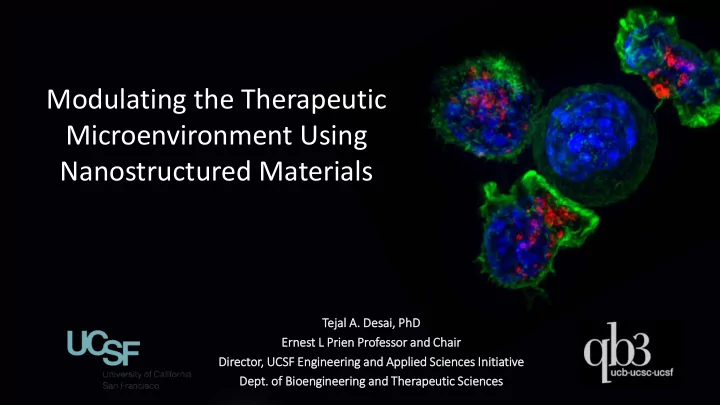
Modulating the Therapeutic Microenvironment Using Nanostructured - - PowerPoint PPT Presentation

modulating the therapeutic microenvironment using
SMART_READER_LITE
LIVE PREVIEW

Modulating the Therapeutic Microenvironment Using Nanostructured - - PowerPoint PPT Presentation

Modulating the Therapeutic Microenvironment Using Nanostructured Materials Tej ejal A l A. Des esai, i, Ph PhD Ernest st L L Pr Prien Professo essor and C nd Chair Direc ector, UCSF E F Engineer eering and nd Appl pplied ed S


slide-1
SLIDE 1

Tej ejal A l A. Des esai, i, Ph PhD Ernest st L L Pr Prien Professo essor and C nd Chair Direc ector, UCSF E F Engineer eering and nd Appl pplied ed S Scien ences I es Initiative

  • Dept. of Bioen

engineer eering a and nd T Ther herape peutic S Scien ences es

Modulating the Therapeutic Microenvironment Using Nanostructured Materials

slide-2
SLIDE 2

Advanced biomaterials for therapeutic delivery

Picture credit: Zhang, Drug Discovery Today

Material Size Surface Shape

CANCER DIABETES HEART DISEASE PSORIASIS ARTHRITIS …

  • Direct biophysical stimulation
  • Biomolecule carrier/presenter
slide-3
SLIDE 3

Micro and Nanostructures Cell –Material Interactions Therapeutic Systems

How can material structure modulate biologic function for therapeutic purposes?

slide-4
SLIDE 4

Desired Routes of Drug Delivery

slide-5
SLIDE 5

Challenges to epithelial drug delivery

5

slide-6
SLIDE 6

Revisiting the small intestine

100 10-2 10-4 10-6 10-8 Length (m)

Small intestine Villi Enterocytes Microvilli

Fox C et al., Journal of Controlled Release, 2015

slide-7
SLIDE 7

Drawbacks of paracellular permeation enhancers

  • Chemical permeation agents

induce opening of tight junctions

  • Examples: Surfactants,

chelators, and toxins

  • Toxicity to cells
  • Non-reversible tight junction
  • pening

Permeation enhancer (red) opens tight junctions for increased drug permeation Non-reversible tight junction damage Membrane damage and cell toxicity

slide-8
SLIDE 8

Opening Epithelial Barriers

Sun et al. Physiological Reviews, 2017.

slide-9
SLIDE 9

Topographical cues can enhance permeation

  • f drug between tight junctions

Nanostructured Film Drug Molecule Cells

Diameter: 200 nm Height: 300 nm Diameter: 800 nm Height: 16 µm High Drug Permeation Low Drug Permeation

Kam et al., Nanoletters, 2014; Stewart et al., Exp. Cell Res, 2017

slide-10
SLIDE 10

Sun, et al. Physiological Reviews, 2017 Walsh, et al. Nano Lett, 2015

Nanostructured microneedles enhance transdermal drug delivery

slide-11
SLIDE 11

200 nm diameter

Fox et al., JCR 2015 Fox et al. ACS Nano 2016

Nanostructured planar particles for enhanced oral delivery

slide-12
SLIDE 12

Nanostructures can be tuned to facilitate permeation

LD100 LD200 LD500 HD100 HD200 HD500

slide-13
SLIDE 13

The process is reversible and involves remodeling of tight junctions

b) a) c)

d

Remove 10 µm 10 µm 10 µm 20 µm 20 µm 20 µm

ZO-1 (tight junction protein), Caco-2 nuclei, F-Actin

slide-14
SLIDE 14

Paracellular or transcellular? Mechanism?

slide-15
SLIDE 15

FITC-IgG present at apical cell-cell borders

slide-16
SLIDE 16

TJ scaffolding protein ZO-1 shows altered morphologies upon NS film treatment

Total level keep the same  reversible

Z-stack-1 Z-stack-2 Z-stack-3 Z-projection

slide-17
SLIDE 17

CRISPR-based gene editing to visualize tight junctions

Endogenous mCherry-ZO1 ZO-1 ICC

Clone isolation

Inserted allele Original allele Inserted allele Original allele

After In Vitro barrier model screening

mCherry ZO-1

Fluorescent protein fusion of TJP-ZO-1

ZO-1 proteins fused with mCherry-reporter at N-terminus through CRISPR

slide-18
SLIDE 18

NS 10-40 mins NS 45-75 mins

Time-Lapse Video of ZO-1 during Nanostructure Treatment (MAX Z-projection, 2 locations 1s=5mins) TEER>>1800

Dynamic changes in TJs with nanostructure contact

slide-19
SLIDE 19

Large aggregates of ZO-1protein are formed on apical side when treated with NS film

slide-20
SLIDE 20

Active interaction between aggregates and border ZO-1

slide-21
SLIDE 21

NS-film treated cells have faster recovery from FRAP

t = 0s t = 100s t = 200s t = 300s Photobleaching in frame

NS-treated Non-treated

slide-22
SLIDE 22

Junctional protein Claudin-4 colocalizes with ZO-1 aggregates

Non-treated Flat NS endogenous mCherry-ZO-1 + AAV exogenous YFP-Cldn3, Z projection of ~1um

Video

slide-23
SLIDE 23

Using physical cues to alter tight junction permeability: implications for delivery

slide-24
SLIDE 24

Harnessing nanotopographical cues for therapy

Smooth Muscle Cells Endothelia l Cells Epithelial Cells Fibroblasts

slide-25
SLIDE 25
  • Dr.
  • r. Xiao H

Huang ng

  • Dr.
  • r. Xiaoy
  • yu Sh

Shi

  • Dr
  • Dr. Anna C

Celli elli

  • Dr. Camer

eron N Nemeth

  • Mike

ke Koval, l, E Emory

  • Thea

ea Mauro, U , UCSF CSF

  • Bo

Bo H Huang, U UCSF CSF

  • NIH

H

  • NSF

SF

  • Kimbe

berl rly C Clark rke

  • Za

Zamb mbone Ltd

  • SPA

PARC

  • Eli L

i Lill lly

Th The The Therapeutic M c Micr cro a and d Nanot

  • tec

echnol

  • logy L

Laboratory at UCSF