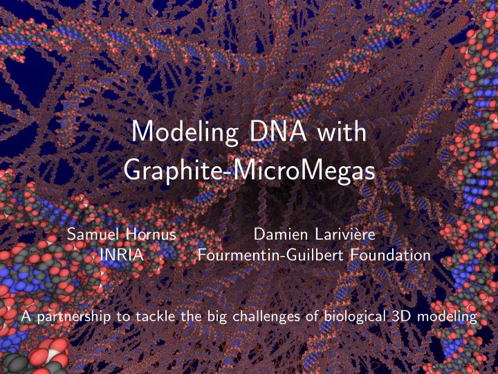
SLIDE 1
Modeling DNA with Graphite-MicroMegas
Samuel Hornus Damien Larivi` ere INRIA Fourmentin-Guilbert Foundation
A partnership to tackle the big challenges of biological 3D modeling
SLIDE 2 Agenda
➲ Biological data deluge ➲ Data mass and comprehension ➲ Comprehension by 3D modeling ➲ Modeling DNA is required ➲ MicroMégas is of great help
SLIDE 3 Biological data deluge
➲ Modern sequencers: One human genome every 14
minutes, 1-2 TB of raw data
A, T, G, C,...
Stuart M. Brown, NYU Langone Medical Center
SLIDE 4 Biological data deluge
➲ Fluorescence
microscopy:
numerous experiments
millions images per run
several tens
Pepperkok & Ellenberg, Nature, 2006
SLIDE 5 Biological data deluge
Julio.Ortiz, Max Planck Institute of Biochemistry
Electron microscopy
SLIDE 6 Data mass and comprehension
➲ 50 years used to dissect cells ➲ Time is come to re- assemble the disconnected
parts
2 µm ~ 3 millions components at the right place
http://www.asiatraveltips.com/newspics/0611/AirbusA380inHK2.jpg David Goodsell, TSRI
SLIDE 7 Comprehension by 3D modeling
➲ 3D modeling is being adopted ➲ 3D software borrowed from “Hollywood” ➲ Need a career time to be learned
Entry of Dengue virus in a cell (Janet Iwasa)
SLIDE 8 Modeling DNA is required
Mycoplasma cell (David Goodsell)
SLIDE 9 Modeling DNA is required
➲ Standard molecular tools and webservices:
- Lack of intuitivity
- Very basic modeling functions
➲ The opensource and intuitive MicroMégas plugin
SLIDE 10 MicroMégas is of great help
➲
The bacterial DNA repair system
Winkler et al, J. Biological Chemistry, March 2011
SLIDE 11 Geometry of DNA
Naive view of DNA is good for low-level modeling:
- string-like structure
- helical shape
- long sequence of very similar “base pairs” (ACGT)
[google image]
SLIDE 12 Geometry of DNA
Computer graphicists translate this structure to:
SLIDE 13 Geometry of DNA
Computer graphicists translate this structure to:
- a curve
- a uniform sampling of orthonormal frames
SLIDE 14 Geometry of DNA
Computer graphicists translate this structure to:
- a curve
- a uniform sampling of orthonormal frames
- instancing of base pairs
SLIDE 15 Geometry of DNA
Computer graphicists translate this structure to:
- a curve
- a uniform sampling of orthonormal frames
- instancing of base pairs
- with twisting: rotation around the tangent vector
SLIDE 16 Modeling a curve
Standard curve models:
ezier curve
ezier curve
- Special case when input is a bare sequence of points
= ⇒ interpolatory subdivision scheme [Dyn, Floater and Hormann 2009]
Modeling a curve
SLIDE 17
Uniform sampling
Generating a uniform sampling with tangent is easy Generating a normal at each sample point is difficult
SLIDE 18
Uniform sampling
Generating a uniform sampling with tangent is easy Generating a normal at each sample point is difficult We want a continuous frame that minimizes torsion E.g. the Fr´ enet-Serret frame is not continuous
SLIDE 19
Uniform sampling
Generating a uniform sampling with tangent is easy Generating a normal at each sample point is difficult We want a continuous frame that minimizes torsion E.g. the Fr´ enet-Serret frame is not continuous Recent technique: very fast and very good approximation: [Rotation Minimizing Frames, ACM ToG 27(1):2, 2008]
SLIDE 20 Visualization with instancing and “ray-casting”
Use OpenGL to instantiate a 3D model of a base-pair in each frame along the curve:
- Setup GL transform matrix
- One call to glDrawArrays to draw one base pair
SLIDE 21 Visualization with instancing and “ray-casting”
A base-pair has ≈ 40 atoms. We setup GLSL programs so that:
- input = array of atoms {center, radius, color}
- geometry shader builds a quad in front of the atom
- pixel shader compute intersection of ray & atom (a sphere)
Use OpenGL to instantiate a 3D model of a base-pair in each frame along the curve:
- Setup GL transform matrix
- One call to glDrawArrays to draw one base pair
SLIDE 22 Visualization with instancing and “ray-casting”
A base-pair has ≈ 40 atoms. We setup GLSL programs so that:
- input = array of atoms {center, radius, color}
- geometry shader builds a quad in front of the atom
- pixel shader compute intersection of ray & atom (a sphere)
Use OpenGL to instantiate a 3D model of a base-pair in each frame along the curve:
- Setup GL transform matrix
- One call to glDrawArrays to draw one base pair
atom camera
SLIDE 23 Visualization with instancing and “ray-casting”
A base-pair has ≈ 40 atoms. We setup GLSL programs so that:
- input = array of atoms {center, radius, color}
- geometry shader builds a quad in front of the atom
- pixel shader compute intersection of ray & atom (a sphere)
Use OpenGL to instantiate a 3D model of a base-pair in each frame along the curve:
- Setup GL transform matrix
- One call to glDrawArrays to draw one base pair
atom camera
SLIDE 24
Level of Details
SLIDE 25
Level of Details
SLIDE 26
Level of Details
SLIDE 27
Level of Details
SLIDE 28 Level of Details
Hierarchy used for
- base-pair picking (on mouseclick)
- LoD selection w.r.t camera position
SLIDE 29
Level of Details
← − Camera is left of screen
SLIDE 30
Thank you
[demo?]
SLIDE 31 Why modeling biological scenes spatially?
Medical illustration
- Popularizing knowledge
- Help scientists’ understanding
- Dynamic simulation
Mre11 David Goodsell c 2010
SLIDE 32 Why modeling biological scenes spatially?
Medical illustration
- Popularizing knowledge
- Help scientists’ understanding
- Dynamic simulation
Mre11 David Goodsell c 2010
Scientific reasons specific to DNA
SLIDE 33
Modeling DNA
Growing importance of modeling and simulation for experiments = ⇒ need for specialized spatial modeling tools for biologists = ⇒ DNA is an important target
SLIDE 34
Modeling DNA
“Modeling DNA in space is such a tedious job!” — microbiologists and illustrators
SLIDE 35 Modeling DNA
“Modeling DNA in space is such a tedious job!” — microbiologists and illustrators Use advanced 3D modeling software
- Maya • 3D Studio • Blender
Or command-line tool and web services with form-based input
- 3DNA • 3D-DART • DNA Maker
And some with “UI”
SLIDE 36 Modeling DNA
“Modeling DNA in space is such a tedious job!” — microbiologists and illustrators Use advanced 3D modeling software
- Maya • 3D Studio • Blender
Or command-line tool and web services with form-based input
- 3DNA • 3D-DART • DNA Maker
And some with “UI”