
SLIDE 1
5/22/2015 1
Germ Cell Tumors of the Testis
Pathology, Immunohistochemistry, and the Often Confusing Appearance of Their Metastases Charles Zaloudek, MD
Department of Pathology UCSF
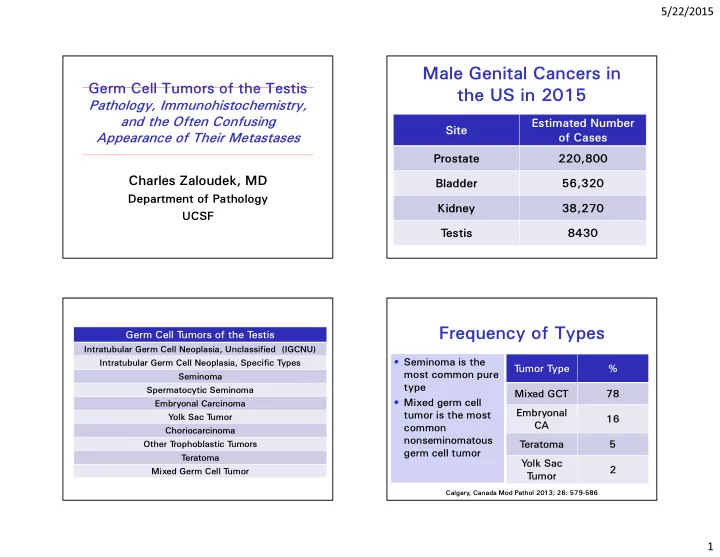
Male Genital Cancers in the US in 2015
Site Estimated Number
- f Cases
Prostate 220,800 Bladder 56,320 Kidney 38,270 T estis 8430
Germ Cell T umors of the T estis
Intratubular Germ Cell Neoplasia, Unclassified (IGCNU) Intratubular Germ Cell Neoplasia, Specific T ypes Seminoma Spermatocytic Seminoma Embryonal Carcinoma Y
- lk Sac T
umor Choriocarcinoma Other T rophoblastic T umors T eratoma Mixed Germ Cell T umor
Frequency of Types
- Seminoma is the
most common pure type
- Mixed germ cell
tumor is the most common nonseminomatous germ cell tumor T umor T ype % Mixed GCT 78 Embryonal CA 16 T eratoma 5 Y
- lk Sac
T umor 2
Calgary , Canada Mod Pathol 2013; 26: 579-586