Lyme neuroborreliosis: infection, immunity, and infl ammation - - PDF document

lyme neuroborreliosis infection immunity and infl ammation
SMART_READER_LITE
LIVE PREVIEW

Lyme neuroborreliosis: infection, immunity, and infl ammation - - PDF document

Review Lyme neuroborreliosis: infection, immunity, and infl ammation Andrew R Pachner, Israel Steiner Lyme neuroborreliosis (LNB), the neurological manifestation of systemic infection with the complex spirochaete Lancet Neurol 2007; 6: 54452


slide-1
SLIDE 1

544 http://neurology.thelancet.com Vol 6 June 2007

Review

Lyme neuroborreliosis: infection, immunity, and infl ammation

Andrew R Pachner, Israel Steiner Lyme neuroborreliosis (LNB), the neurological manifestation of systemic infection with the complex spirochaete Borrelia burgdorferi, can pose a challenge for practising neurologists. This Review is a summary of clinical presentation, diagnosis, and therapy, as well as of recent advances in our understanding of LNB. Many new insights have been gained through work in experimental models of the disease. An appreciation of the genetic heterogeneity of the causative pathogen has helped clinicians in their understanding of the diverse presentations of LNB.

Neuroborreliosis and Lyme disease

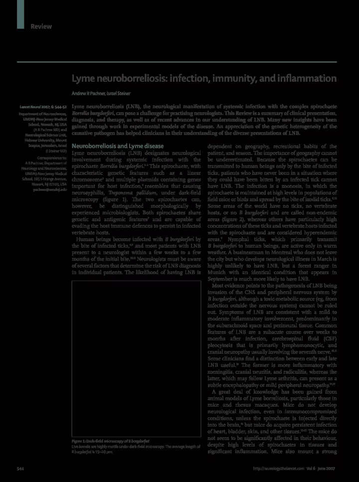
Lyme neuroborreliosis (LNB) designates neurological involvement during systemic infection with the spirochaete Borrelia burgdorferi.1–4 This spirochaete, with characteristic genetic features such as a linear chromosome5 and multiple plasmids containing genes important for host infection,6 resembles that causing neurosyphilis, Treponema pallidum, under dark-fi eld microscopy (fi gure 1). The two spirochaetes can, however, be distinguished morphologically by experienced microbiologists. Both spirochaetes share genetic and antigenic features7 and are capable of evading the host immune defences to persist in infected vertebrate hosts. Human beings become infected with B burgdorferi by the bite of infected ticks,8,9 and most patients with LNB present to a neurologist within a few weeks to a few months of the initial bite.10,11 Neurologists must be aware

  • f several factors that determine the risk of LNB diagnosis

in individual patients. The likelihood of having LNB is dependent on geography, recreational habits of the patient, and season. The importance of geography cannot be underestimated. Because the spirochaetes can be transmitted to human beings only by the bite of infected ticks, patients who have never been in a situation where they could have been bitten by an infected tick cannot have LNB. The infection is a zoonosis, in which the spirochaete is maintained at high levels in populations of fi eld mice or birds and spread by the bite of ixodid ticks.8,12 Some areas of the world have no ticks, no vertebrate hosts, or no B burgdorferi and are called non-endemic areas (fi gure 2), whereas others have particularly high concentrations of these ticks and vertebrate hosts infected with the spirochaete and are considered hyperendemic areas.9 Nymphal ticks, which primarily transmit B burgdorferi to human beings, are active only in warm

  • weather. A businessman in Montreal who does not leave

the city but who develops neurological illness in March is highly unlikely to have LNB, but a forest ranger in Munich with an identical condition that appears in September is much more likely to have LNB. Most evidence points to the pathogenesis of LNB being invasion of the CNS and peripheral nervous system by B burgdorferi, although a toxic-metabolic source (eg, from infection outside the nervous system) cannot be ruled

  • ut. Symptoms of LNB are consistent with a mild to

moderate infl ammatory involvement, predominantly in the subarachnoid space and perineural tissue. Common features of LNB are a subacute course over weeks to months after infection, cerebrospinal fl uid (CSF) pleocytosis that is primarily lymphomonocytic, and cranial neuropathy usually involving the seventh nerve.10,11 Some clinicians fi nd a distinction between early and late LNB useful.13 The former is more infl ammatory with meningitis, cranial neuritis, and radiculitis, whereas the latter, which may follow Lyme arthritis, can present as a subtle encephalopathy or mild peripheral neuropathy.14,15 A great deal of knowledge has been gained from animal models of Lyme borreliosis, particularly those in mice and rhesus macaques. Mice do not develop neurological infection, even in immunocompromised conditions, unless the spirochaete is injected directly into the brain,16 but mice do acquire persistent infection

  • f heart, bladder, skin, and other tissues.17–21 The mice do

not seem to be signifi cantly aff ected in their behaviour, despite high levels of spirochaetes in tissues and signifi cant infl

  • ammation. Mice also mount a strong

Lancet Neurol 2007; 6: 544–52 Department of Neurosciences, UMDNJ-New Jersey Medical School, Newark, NJ, USA (A R Pachner MD) and Neurological Science Unit, Hebrew University, Mount Scopus, Jerusalem, Israel (I Steiner MD) Correspondence to: A R Pachner, Department of Neurology and Neurosciences, UMDNJ-New Jersey Medical School, 185 S Orange Avenue, Newark, NJ 07103, USA pachner@umdnj.edu

Figure 1: Dark-fi eld microscopy of B burgdorferi Live borrelia are highly motile under dark-fi eld microscopy. The average length of B burgdorferi is 15–20 μm.

slide-2
SLIDE 2

http://neurology.thelancet.com Vol 6 June 2007 545

Review

humoral and cellular immune response, which is ineff ective in clearing the spirochaete. The human and non-human primate immune response is much more eff ective against the spirochaete. However, when mild immunosuppression is induced in rhesus macaques, the spirochaete establishes infection22,23 with nests of spirochaetes surrounded by infl ammatory cells in many

  • rgans, including the CNS and peripheral nervous

system.24–28 Infl ammation in the nervous system in rhesus macaques is primarily localised to nerve roots, dorsal root ganglia, and leptomeninges. T cells and plasma cells are the predominant infl ammatory cells. Signifi cantly increased amounts of IgG, IgM, and C1q are found in infl amed spinal cords. Spirochaetes can be visualised by immunohistochemistry24 in the leptomeninges, nerve roots, and dorsal root ganglia, but not in the CNS parenchyma (fi gure 3). These data are consistent with the pattern of clinical involvement in infected human beings, with meningitis and radiculitis predominating, and parenchymal involvement occurring

  • nly rarely.

Epidemiology

There are substantial clinical diff erences in LNB depending on whether infection occurred in the USA or Europe (table), because of genetic diff erences between the strain that causes all cases of US LNB, B burgdorferi sensu stricto, and the European strains B garinii and B afzelii;29,30 B burgdorferi sensu stricto has been isolated from a small percentage of European patients but almost never causes LNB in Europe. Diff erent disease manifestations were induced when B burgdorferi sensu stricto spirochaetes isolated from the CSF of American patients and B garinii or B afzelii from European patients were injected into mice.31 There are also diff erences in the life cycle of B burgdorferi in the various continents. The tick Ixodes scapularis, which primarily feeds on fi eld mice but may also be maintained in other rodents, is the main host for the spirochaete in the USA, whereas in Europe I ricinus feeds on several animals, but may become infected with B garinii, mostly from birds;32 in addition, the seabird tick I uriae may be important for B garinii infection in Europe, especially in the North Atlantic.33 LNB probably also occurs in Asia, and is under active investigation in China, Japan, and Korea. Russian LNB has been well documented.34 Lyme disease may not exist in South America35 or Africa,36 and there are many areas

  • f Asia and even Europe and North America that have no

Lyme disease because of the absence of the spirochaetes in ticks or of the vertebrate hosts. Consistent with the complex epidemiology is the great degree of genetic heterogeneity among the three main species. Further complicating the picture is the fact that ticks may also carry other infectious organisms such as Babesia and Ehrlichia, and coinfections, although uncommon, are possible.

Diagnosis

Diagnosis of LNB would ideally be made by demonstration of the causative pathogen in the CSF. Culture37 or PCR38 of CSF samples have been used for this purpose, and spirochaetes can readily be detected by PCR39–41 or histology19,24 in tissues of experimentally infected animals. The yield of CSF culture in patients with LNB is less than 5%; CSF PCR has a higher

  • sensitivity. While it can be positive in up to 40% of

patients with LNB, especially in patients with meningitis,38 this is usually early in the course of

  • infection. Unfortunately, the spirochaete is primarily a

tissue-based organism, and does not appear for extended durations in blood or CSF. Consequently, direct demonstration of the spirochaete in blood or CSF is impractical in routine clinical settings because of its low

  • yield. Thus, the diagnosis must rely on a combination of

history, examination, routine analyses of CSF, and antibody studies of serum and CSF.42,43

Figure 2: World-wide distribution of ixodid ticks capable of carrying and transmitting B burgdorferi to humans Reproduced with permission of WHO. Figure 3: B burgdorferi in the dorsal root ganglia of Rhesus macaques during chronic experimental infection Spirochaetes have been labelled by immunohistochemistry with a high-titre rabbit polyclonal antiserum. The average length of B burgdorferi is 15–20 μm.

slide-3
SLIDE 3

546 http://neurology.thelancet.com Vol 6 June 2007

Review

History

Exposure As mentioned, geography and outdoor exposure are critical factors. Neurologists in non-endemic areas should

  • btain a travel history from patients who might have

travelled to an endemic area and been infected there. Infected ticks are usually present in suburbs or countryside, especially where there are many deer, although it is theoretically possible to be infected in city parks.44 However, an individual who stays in the city and does not venture out is very unlikely to develop LNB. Because the ixodid ticks are small and inconspicuous, most patients do not recall the tick bite that resulted in

  • LNB. Thus, asking the patient about tick bites is not

necessarily a helpful addition to the history. Erythema migrans Erythema migrans, which is a diagnostic hallmark of the disease, is a chronic area of expanding erythema greater than 5 cm in diameter, commonly raised and sometimes

  • itchy. This circular or elliptical red area that spreads

centrifugally daily (fi gure 4) is caused by movement of spirochaetes through the skin. A history of erythema migrans is very common in LNB, and should be actively

  • explored. Because LNB typically occurs within the fi

rst few months of infection and erythema migrans is an early manifestation, usually in the fi rst few weeks, the rash is commonly recalled by patients. LNB in the USA usually occurs after the erythema migrans rash. In the placebo group of a large vaccine study involving more than 5000 patients in each arm45 in areas of northeast USA that are endemic in Lyme disease, there were only two patients who developed LNB de novo without an earlier manifestation of the systemic disease including erythema migrans. In this study, neurological involvement occurred in only 3% of the 86 patients with Lyme borreliosis. By contrast, 81 of the 86 patients (94%) who developed Lyme borreliosis had eyrthema migrans as their manifestation. The situation is very diff erent with the European forms

  • f LNB. In Bannwarth’s syndrome in Europe, erythema

migrans can occur, but is a preceding event in only a few

  • cases. In a study of 404 patients of Bannwarth’s syndrome

in Germany,46 most cases of LNB (58%) occurred without a preceding erythema migrans. In this study, LNB was the presentation of Lyme disease in 49% of cases and was the most common manifestation, and erythema migrans, arthritis, carditis, and acrodermatitis were less common. Thus, many patients infected in Europe will not have erythema migrans before their LNB, which makes it important to obtain a history of foreign travel. Nevertheless, now that erythema migrans is generally recognised and treated aggressively in the USA, the incidence of LNB without this preceding symptom will probably be higher, although no studies have addressed this issue. Erythema migrans also aff ects children with Lyme

  • disease. In a large study of pediatric Lyme disease in the

USA,47 the spectrum of the disease was similar to that of adult LNB, with 90% of children presenting with erythema migrans, and only 5% had either facial palsy or meningitis; smaller studies have noted a higher incidence of LNB in children than in adults.48 As with European adults, the incidence of neurological involvement relative to other disease manifestations is high in children in Europe. Symptoms of LNB American LNB almost always presents as a subacute meningitis without or with associated facial palsy within a few weeks to a few months of infection or the erythema migrans rash.11,42 Headache, meningismus, numbness and tingling that is not attributable to a defi nite anatomical localisation, myalgia, fatigue, and malaise are all common. Patients may have mild cognitive symptoms but do not have severe organic brain syndromes. Although radicular symptoms may be present, painful radiculitis is uncommon. European LNB usually presents as Bannwarth’s syndrome10,49 (also called Garin-Bujadoux syndrome).50 Bannwarth’s syndrome is more easily identifi ed because

  • f its presentation as painful radiculitis.51 86% of LNB

cases in Europe present with painful radiculitis with or without associated paresis. This is of course not pathognomonic and European neurologists have been warned that not all painful radiculitides are caused by spirochaetes.52 The pain of Bannwarth’s syndrome is frequently chronic, lasting for weeks to months after initial infection; it may be severe, commonly worsening at night, and is often described as lancinating. Bannwarth’s syndrome is also sometimes called “lymphocytic meningoradiculitis”,53 because

  • f

accompanying meningitis and the lymphocytosis in the spinal fl

  • uid. Some clinicians feel that Bannwarth’s

syndrome may be a unique clinical hallmark of LNB caused by B garinii,54 but Bannwarth’s syndrome can

  • ccasionally be seen in American LNB, caused by

B burgdorferi sensu stricto.55

Clinical feature American LNB European LNB Causative Borrelia subspecies B burgdorferi sensu stricto Mostly B garinii, occasionally B afzelii LNB as a percentage of all Lyme cases <10% >35% Multiple erythema migrans lesions Common Uncommon Painful radiculitis Rare (<10%) Common (>50%) “Aseptic” meningitis presentation Majority Minority Cranial nerve involvement VII, very rarely others Usually VII, but can include others Associated chronic skin manifestation (lymphocytoma or ACA) Never Not rare Associated with Lyme arthritis Common Almost never Chronic encephalomyeloradiculitis Very rare (<0.1% of LNB) More frequent, but unusual (<3% of LNB) Intrathecal antibody production Minority of cases Common (>50%) ACA=acrodermatitis chronica atrophicans. Table: Diff erences between European and American LNB

slide-4
SLIDE 4

http://neurology.thelancet.com Vol 6 June 2007 547

Review

In both forms of LNB, peripheral neuropathy, as distinct from facial neuropathy, can occur with symptoms

  • f numbness and tingling. The neuropathy is generally

not severe, and disabling weakness is unusual. Diff erentiation between the radiculitis of Bannwarth’s syndrome and neuritis is clinically impossible in some

  • cases. Most neuropathies from Lyme disease are axonal,56

especially those associated with acrodermatitis chronicum atrophicans,57 a chronic skin lesion primarily caused by north European strains of B burgdorferi.

Signs

There are no fi ndings on neurological examination that are absolutely specifi c for LNB. Some patients have bilateral facial palsy in the summer or autumn, a fi nding that, in endemic areas, is highly predictive of LNB. Examination should also include the dermatological search for erythema

  • migrans. The combination of headache, facial palsy, and a

characteristic erythema migrans in areas endemic for Lyme disease in the summer or autumn is suffi cient for a

  • diagnosis. A search of the skin for coexisting erythema

migrans in patients with chronic headache in an area endemic for Lyme disease may be rewarding. Unfortunately, erythema migrans is usually not present when patients with LNB present to physicians,58 and suspicion of LNB needs to be confi rmed by looking for antibodies for spirochaetes in serum59 and CSF.60

Laboratory studies

CSF or serum antibody index The gold standard for the laboratory diagnosis of bacterial infections is identifi cation of the pathogen. Unfortunately, in LNB, as in neurosyphilis, the yield of PCR or culture is too low. However, LNB usually presents at a time when a strong immune response to the spirochaetes is mounted, resulting in high titres of specifi c antibodies for B burgdorferi in serum and CSF. In European LNB, the antibody index is typically used to aid the diagnosis of LNB:61 specifi c antibody titres are measured in the CSF and in the serum and are calculated to measure intrathecal synthesis of antibodies for spirochaetes. An antibody index above 1 means that the antibody is at a higher relative level in the CSF than in the serum.62 Lumbar puncture in patients with LNB will also generally support the diagnosis by showing a monocytic or lymphocytic pleocytosis, commonly with a mild to moderately high protein concentration, consistent with an infl ammatory process.63 Imaging studies are generally not helpful,64

A B C D

Figure 4: Erythema migrans in an infant during the summer in a highly endemic area (New Jersey, USA) Day 1: the rash was fi rst identifi ed on this day; note the “bulls-eye” appearance, which is commonly, but not universally present (A). Day 5: the rash has spread centrifugally, and there is relative central clearing (B). Day 6: more spread (C). Day 9: 3 days after antibiotic therapy, the rash has almost completely cleared (D).

slide-5
SLIDE 5

548 http://neurology.thelancet.com Vol 6 June 2007

Review

except to rule out other neurological processes, although

  • ccasionally the subarachnoid infl

ammation will be profound enough for gadolinium enhancement of the

  • meninges. SPECT scanning has been used by some

clinicians, but its reliability in LNB is unproven. In American LNB, the CSF antibody index is not commonly used, possibly because there has not been an extensive study to assess its usefulness in the American LNB setting relative to the usefulness of serum antibodies in patients with possible LNB. Using an antibody capture technique, Steere and colleagues65 showed signifi cant diff erences between American and European patients with LNB, indicating that the sensitivity of the CSF antibody-index assay for American LNB may not be high relative to European LNB. Thus, at this time, there are no well-accepted criteria for the diagnosis of American LNB, and it is reasonable to look for antibodies for B burgdorferi in serum for diagnosis with the approach outlined below. Two-step approach for serum antibody assays Patients with American LNB are infected with strains of B burgdorferi sensu stricto that are relatively homogeneous compared to the situation in Europe, where there is great genetic heterogeneity among the strains that infect human

  • beings. Thus, immune responses in American patients are

directed to a predictable medley of antigens, and antibody testing is reasonably straightforward. In 1994, the American Centers for Disease Control in association with the Association of State and Territorial Public Health Laboratory Directors recommended a two-step approach to testing in which the fi rst step was an ELISA. If the ELISA— which is fast, inexpensive, and highly sensitive—is positive, the serum sample is further tested in an immunoblot assay (a western blot).66 The western blot, using the so-called Dressler criteria,67 adds the specifi city missing in the ELISA

  • assay. The ELISA–western blot combination has been

validated both in formal studies68 and in clinical experience. Although in extensive use, this technique suff ers from the same problems as any serological assay: it is not highly sensitive for very early Lyme borreliosis, and it cannot readily distinguish between active infection and previous

  • exposure. Nevertheless, the assay is very useful for LNB,

because neurological involvement presents weeks to months after initial infection.11,58 Thus, a negative serum assay for antibodies for B burgdorferi in the USA will generally rule out LNB. Because the strains of B burgdorferi causing LNB in Europe are much more heterogeneous, the ELISA–western blotting of serum becomes more complicated and less reliable,69 and the antibody index remains the mainstay of laboratory dignosis in Europe. The development of recombinant Borrelia proteins for immunodiagnosis in European Lyme disease research centres70,71 may help address this problem because in animal models, Borrelia recombinant proteins have proven very helpful in defi ning the humoral response.23 The recent development of an assay for serum antibodies to the peptide from the sixth invariant region of the VlsE lipoprotein of B burgdorferi (see below) may pave the way for the development of a more useful assay in serum for LNB in Europe.72 Overlap with other neurological diseases Despite the combination of clinical setting and serological confi rmation, the diagnosis of LNB can sometimes be less than straightforward. The diagnosis is sometimes considered in patients with multiple sclerosis,73,74 although Lyme disease rarely presents with evidence of focal CNS damage, or a clearly abnormal MRI;75 non-specifi c small white-matter abnormalities on brain MRI can be seen in a few patients76 and there is a report of gadolinium-enhanced cranial nerves in a patient with European LNB.77 Sometimes, an MRI brain scan in a patient with seropositivity can be abnormal because patients with multiple sclerosis can be seropositive from previous exposure to the spirochaete.78 Fatigue, myalgia, and arthralgia can be major symptoms of any chronic infection, and occasionally LNB may be considered in patients with chronic fatigue syndrome79 or fi bromyalgia.80,81 Fatigue, malaise, and headache were common symptoms of LNB in chronic LNB caused by B afzelii in a series reported by Strle and co-workers.54 These patients had milder CSF abnormalities than those with LNB caused by B garinii. Active encephalitis in LNB is highly unusual, but encephalopathy has been reported.82–85 This encephalopathy, which is an uncommon manifestation of LNB, generally consists of symptoms of malaise and diffi culties with concentration, sometimes with defi cits of verbal memory, mental agility, and verbal functions; problem solving skills, mental speed, and visuospatial functions are commonly unimpaired.85 Associated fatigue and arthralgias are common with the encephalopathy. This encephalopathy can also be seen in children.86 The overlap with fi bromyalgia is evident, although fi bromyalgia with its disabling pain and trigger points can usually be diff erentiated from LNB. The encephalopathy has generally been seen in patients in whom the infection has proceeded for months to years without previous treatment. The pathogenesis is unclear, although the fact that about two-thirds of patients with it respond to 2 weeks of intravenous antibiotics indicates that it may be secondary to persistent infection.

Therapy

The natural history and ultimate prognosis of untreated LNB is unknown, but in many patients the infection can probably resolve without antibiotics. However, antibiotics are given to hasten clearance of the infection, speed resolution of symptoms, and prevent the development of late manifestations of the disease such as arthritis and acrodermatitis chronica atrophicans. The antibiotic treatment of choice for LNB in the USA at this time is an intravenous cephalosporin,87 such as ceftriaxone, or penicillin for 2–4 weeks. In the fi rst study of antibiotic therapy for Lyme neuroborreliosis, it was found that penicillin G, 20 million units/day in divided doses for

slide-6
SLIDE 6

http://neurology.thelancet.com Vol 6 June 2007 549

Review

10 days was successful.88 Others have used ceftriaxone, which is thought to be better in view of its long half-life, resulting in maintenance of high concentrations in serum for a longer period of time, and its ability to penetrate the blood–brain barrier readily and maintain high CSF concentrations.89 The dose of ceftriaxone is generally intravenous or intramuscular 1–2 g twice a day for 14–28 days.13,90,91 In a large European study, oral doxycycline was as eff ective as intravenous antibiotics in treating LNB. The dose of doxycycline used was 200 mg orally daily for 14 days.92,93 Thus, oral therapy is a reasonable alternative to intravenous agents in neurological involvement in this infection.91 Therapy is highly successful, and residual symptoms after therapy have been found predominantly in patients who had irreversible damage to the facial nerve

  • r nerve roots before therapy.94 However, treatment failures

have been seen in response to both doxycycline and ceftriaxone therapy and it is reasonable to try additional therapy with an alternative antibiotic under those circumstances. Lyme disease is a highly infl ammatory disease in many

  • cases. In fact one of the mysteries of the infection is how

a few spirochaetes are able to provoke such intense infl

  • ammation. Some of the symptoms are secondary to

the infl ammation induced by the spirochaete, not by the spirochaete load. Thus, eff ective therapy includes minimisation of infl ammation as well as eradication of

  • infection. In our experience, non-steroidal anti-

infl ammatory drugs can be eff ective in improving symptoms of arthralgias, myalgias, and headache in selected patients with active symptoms of LNB. Because LNB responds relatively quickly to antibiotics, the need, if there is one, for non-steroidal anti-infl ammatory drugs is usually for short periods. Corticosteroids have been used by some clinicians to treat infl ammatory syndromes in Lyme disease.15 Corticosteroids may interfere with immune-mediated killing of spirochaetes, and thus should not be given unless a patient has undergone an adequate period of antibiotics to control the infection. Corticosteroids may be indicated in a small subset of patients who have infl ammatory syndromes that persist after antibiotics and do not respond to non-steroidal anti-infl ammatory drugs,95 but they are not considered standard therapy in patients with Lyme borreliosis. Neurologists might occasionally be faced with idiopathic facial palsies for which they are considering corticosteroid therapy, but there is a possibility that the facial palsy may be due to infection with B burgdorferi. For such patients, a positive Lyme serology or history of erythema migrans will tip the scales towards LNB. Alternatively, absence of exposure in an endemic area, or presentation after months

  • f winter weather will suggest Bell’s palsy, because

B burgdorferi infection is rare in the winter months. In those situations in which diff erentiation is diffi cult, neurologists have many options for both diagnosis and therapy. Obtaining a Lyme antibody assay is defi nitely indicated, because if Lyme disease is present, it should be treated. If the results of the Lyme antibody assay are negative, it should be repeated after 2–4 weeks, because facial palsy can be an early manifestation of infection before seroconversion; neurologists should also be aware that if steroid treatment for Lyme disease is chosen, it can delay seroconversion during infection. The therapeutic decisions are even further complicated by the facts that antibiotics do not aff ect the natural history of the facial palsy in LNB but do prevent late manifestations such as arthritis,96 that corticosteroids are not universally accepted as being eff ective therapy in Bell’s palsy, and that steroids increase spirochaetal load in the rhesus macaque model of LNB.22 These considerations dictate that therapeutic decisions on patients with facial palsy need to be made on a case-by-case basis.

Post-Lyme disease syndrome

Most patients with LNB who received adequate antibiotic therapy will have a prompt recovery, with complete resolution of symptoms within a few weeks to months. However, some patients continue to have prolonged symptoms such as fatigue, myalgias, arthralgias, low-grade cognitive diffi culties, and sleep problems despite therapy,97 and these patients are sometimes diagnosed as having post-Lyme disease98 and may come to the attention of

  • neurologists. The pathogenesis of post-Lyme disease is

unknown, but could be due to persistence of the spirochaete after antibiotics, a situation that has been documented, using PCR but not culture, in some animal models.99,100 However, the results of two careful clinical trials have provided strong evidence against this possibility in human beings.101 Alternatively, symptoms may be due to the lack of complete clearance of Borrelia glycolipids that are highly infl ammatory.102–104 The optimum pathogenesis and management of post-Lyme syndrome remain controversial, although most clinicians treat it symptomatically; fortunately, it is uncommon.

CXCL13—a new biomarker for LNB

Given the absence of direct diagnosis of pathogen in the CSF either by PCR or culture, and the vagaries of serological measures, it would be ideal to have a biomarker for the disease. In 2002, we reported that the chemokine CXCL13 was highly expressed in the tissue of infected rhesus macaques and that CXCL13 correlated well with the presence of spirochaetes.28,105 We subsequently showed that this was probably related to direct spirochaete stimulation of dendritic cells.106 Rupprecht and co-workers107,108 then described the presence of CXCL13 in the CSF of all patients with Bannwarth’s syndrome and its absence in all patients with other diagnoses. Chemokines are signalling molecules for leucocyte migration, and CXCL13 is primarily a B-cell-tropic molecule.109 The fi ndings are consistent with the fact that the humoral component of the immune response is primarily involved in spirochaete killing.110

slide-7
SLIDE 7

550 http://neurology.thelancet.com Vol 6 June 2007

Review

Host evasion and complement inhibition

The inability of murine antibodies to clear the spirochaete is probably due to active mechanisms of immune evasion by the spirochaete, particularly the production of proteins that bind to and inactivate mouse complement, markedly decreasing the destructive capacity of antibodies against the spirochaete.111,112 Molecules produced by pathogens that specifi cally target the immune system of the host to allow persistent infection are known as immunoevasins; com- plement inactivators are likely to be only one of several immun

  • evasins produced by B burgdorferi. Mice serve as a

reservoir for the infection in the wild, so it is not surprising that such mechanisms exist to allow persistence at high

  • levels. However, primates, such as human beings and

rhesus macaques, are dead-end hosts for B burgdorferi (ie, they do not transmit the infection to ticks). Hence, most Borrelia strains generally do not have mechanisms to evade primate complement, and infection results in rapid rises in antibodies for the spirochaetes, with unimpeded complement acitivity and clearance of the spirochaete in most animals without the necessity for antibiotics.27,113,114 Some B garinii strains may represent exceptions to this rule; a recent study115 documented that neuroinvasive B garinii strains express complement factor H-binding proteins.

Improvement in diagnosis using serology—the C6 assay

Although the two-step technique of ELISA followed by western blot has been very successful in assisting neurologists in the diagnosis of LNB, a one-step, less expensive technique, would be preferred. Recent studies116,117 have confi rmed that a new technique, called the C6 ELISA, is a promising candidate to replace the two-step testing. This ELISA takes advantage of the conserved sequence of this protein and its nearly universal expression. C6 is a portion of the vls (variable major protein-like sequence) E locus of B burgderfori,118 which encodes an antigenic variation system that is related to the VMP system of relapsing fever borreliae,119,120 and uses a combinatorial strategy of gene cassettes. The assay remains experimental and should not be used as a replacement for the two-step technique; studies are ongoing.

Conclusion

Great strides have been made over the past two decades in understanding LNB since its fi rst description almost 30 years ago as a manifestation of disseminated Lyme disease3 and since its fi rst description as a clinical entity 85 years ago.50 The pathogenesis is to a great degree understood, the diagnosis has become straightforward with potentially improved diagnostic assays on the horizon, biomarkers with great promise are being developed, therapy has been defi ned, and the prognosis

  • f the treated infection well-characterised. Given its lack
  • f a distinctive and unique clinical presentation,

neurologists can occasionally fi nd recognition of this infection diffi cult, but should be confi dent in the currently available diagnostic assays and antibiotics.

Contributions ARP was responsible for the literature search and for writing most sections of the Review. IS was responsible for writing some sections and editing the Review. Confl icts of interest We have no confl icts of interest. References 1 Burgdorfer W, Barbour AG, Hayes SF, Benach JL, Grunwaldt E, Davis JP. Lyme disease-a tick-borne spirochetosis? Science 1982; 216: 1317–19. 2 Steere AC. Lyme disease. N Engl J Med 2001; 345: 115–25. 3 Reik L, Steere AC, Bartenhagen NH, Shope RE, Malawista SE. Neurologic abnormalities of Lyme disease. Medicine 1979; 58: 281–94. 4 Pachner AR, Steere AC. Neurological fi ndings of Lyme disease. Yale J Biol Med 1984; 57: 481–83. 5 Ferdows MS, Barbour AG. Megabase-sized linear DNA in the bacterium Borrelia burgdorferi, the Lyme disease agent. Proc Natl Acad Sci USA 1989; 86: 5969–73. 6 Barbour AG. The molecular biology of Borrelia. Rev Infect Dis 1989; 11 (suppl 6): S1470–74. 7 Magnarelli LA, Anderson JF, Johnson RC. Cross-reactivity in serological tests for Lyme disease and other spirochetal infections. J Infect Dis 1987; 156: 183–88. 8 Fish D. Environmental risk and prevention of Lyme disease. Am J Med 1995; 98: 2S–8S. 9 Lastavica CC, Wilson ML, Berardi VP, Spielman A, Deblinger RD. Rapid emergence of a focal epidemic of Lyme disease in coastal

  • Massachusetts. N Engl J Med 1989; 320: 133–37

. 10 Ackermann R, Horstrup P, Schmidt R. Tick-borne meningopoly- neuritis (Garin-Bujadoux, Bannwarth). Yale J Biol Med 1984; 57: 485–90. 11 Pachner AR, Steere AC. The triad of neurologic manifestations of Lyme disease: meningitis, cranial neuritis, and radiculoneuritis. Neurology 1985; 35: 47–53. 12 Magnarelli LA, Anderson JF, Hyland KE, Fish D, Mcaninch JB. Serologic analyses of Peromyscus leucopus, a rodent reservoir for Borrelia burgdorferi, in northeastern United States. J Clin Microbiol 1988; 26: 1138–41. 13 Logigian EL, Kaplan RF, Steere AC. Chronic neurologic manifestations

  • f Lyme disease [see comments]. N Engl J Med 1990; 323: 1438–44.

14 Halperin JJ. Neuroborreliosis. Am J Med 1995; 98: 52S–6S. 15 Kaiser R. Neuroborreliosis. J Neurol 1998; 245: 247–55. 16 Li L, Narayan K, Pak E, Pachner A. Intrathecal antibody production in a mouse model of Lyme neuroborreliosis. J Neuroimmunol 2006; 173: 56–68. 17 Pachner AR, Delaney E, Ricalton NS. Murine Lyme borreliosis: route of inoculation determines immune response and infectivity. Reg Immunol 1992; 4: 345–51. 18 Pachner AR, Itano A. Borrelia burgdorferi infection of the brain: characterization of the organism and response to antibiotics and immune sera in the mouse model. Neurology 1990; 40: 1535–40. 19 Pachner AR, Basta J, Delaney E, Hulinska D. Localization of Borrelia burgdorferi in murine Lyme borreliosis by electron

  • microscopy. Am J Trop Med Hyg 1995; 52: 128–33.

20 Barthold SW, deSouza MS, Janotka JL, Smith AL, Persing DH. Chronic Lyme borreliosis in the laboratory mouse. Am J Pathol 1993; 143: 959–72.

Search strategy and selection criteria References were identifi ed through searches of PubMed from January 1975 to March 2007 with the terms “Lyme”, “neuroborreliosis”, “brain”, “spinal cord”, “nerve”, “radiculopathy”, “encephalopathy”, “Borrelia”, “CSF”, “therapy”, and “facial” and through searches of the authors’

  • wn fi
  • les. Only papers written in English were reviewed.
slide-8
SLIDE 8

http://neurology.thelancet.com Vol 6 June 2007 551

Review

21 Barthold SW, de Souza MS, Janotka JL, Smith AL, Persing DH. Chronic Lyme borreliosis in the laboratory mouse. Am J Pathol 1993; 143: 959–71. 22 Pachner AR, Amemiya K, Bartlett M, Schaefer H, Reddy K, Zhang WF. Lyme borreliosis in rhesus macaques: eff ects of corticosteroids on spirochetal load and isotype switching of anti- Borrelia burgdorferi antibody. Clin Diagn Lab Immunol 2001; 8: 225–32. 23 Pachner AR, Dail D, Li L, et al. Humoral immune response associated with lyme borreliosis in nonhuman primates: analysis by immunoblotting and enzyme-linked immunosorbent assay with sonicates or recombinant proteins. Clin Diagn Lab Immunol 2002; 9: 1348–55. 24 Cadavid D, O’Neill T, Schaefer H, Pachner AR. Localization of Borrelia burgdorferi in the nervous system and other organs in a nonhuman primate model of lyme disease. Lab Invest 2000; 80: 1043–54. 25 Bai Y, Narayan K, Dail D, Sondey M, Hodzic E, Barthold SW, et al. Spinal cord involvement in the nonhuman primate model of Lyme

  • disease. Lab Invest 2004; 84: 160–72.

26 Pachner AR, Cadavid D, Shu G, et al. Central and peripheral nervous system infection, immunity, and infl ammation in the NHP model of Lyme borreliosis. Ann Neurol 2001; 50: 330–38. 27 Cadavid D, Bai Y, Dail D, et al. Infection and infl ammation in skeletal muscle from nonhuman primates infected with diff erent genospecies of the Lyme disease spirochete Borrelia burgdorferi. Infect Immun 2003; 71: 7087–98. 28 Cadavid D, Bai Y, Hodzic E, Narayan K, Barthold SW, Pachner AR. Cardiac involvement in non-human primates infected with the Lyme disease spirochete Borrelia burgdorferi. Lab Invest 2004; 84: 1439–50. 29 Wang G, van Dam AP, Schwartz I, Dankert J. Molecular typing of Borrelia burgdorferi sensu lato: taxonomic, epidemiological, and clinical implications. Clin Microbiol Rev 1999; 12: 633–53. 30 Glockner G, Lehmann R, Romualdi A, et al. Comparative analysis

  • f the Borrelia garinii genome. Nucleic Acids Res 2004; 32: 6038–46.

31 Pachner AR, Dail D, Bai Y, et al. Genotype determines phenotype in experimental Lyme borreliosis. Ann Neurol 2004; 56: 361–70. 32 van Dam AP. Diversity of Ixodes-borne Borrelia species—clinical, pathogenetic, and diagnostic implications and impact on vaccine

  • development. Vector Borne Zoonotic Dis 2002; 2: 249–54.

33 Bunikis J, Olsen B, Fingerle V, Bonnedahl J, Wilske B, Bergstrom S. Molecular polymorphism of the lyme disease agent Borrelia garinii in northern Europe is infl uenced by a novel enzootic Borrelia focus in the North Atlantic. J Clin Microbiol 1996; 34: 364–68. 34 Lesnik OM, Istomina O, Rijpkema S, Bruininck H, Beliaeva ML. The clinical manifestations of Lyme borreliosis in the Middle Urals and their association with Borrelia burgdorferi genospecies. Ter Arkh 1997; 69: 9–12. 35 Mattar S, Lopez Valencia G. Searching for Lyme disease in Colombia: a preliminary study on the vector. J Med Entomol 1998; 35: 324–26. 36 Collares-Pereira M, Gomes AC, Prassad M, et al. Preliminary survey

  • f leptospirosis and Lyme disease amongst febrile patients

attending community hospital ambulatory care in Maputo,

  • Mozambique. Cent Afr J Med 1997; 43: 234–38.

37 Karlsson M, Hovind-Hougen K, Svenungsson B, Stiernstedt G. Cultivation and characterization of spirochetes from cerebrospinal fl uid of patients with Lyme borreliosis. J Clin Microbiol 1990; 28: 473–79. 38 Pachner AR, Delaney E. The polymerase chain reaction (PCR) in the diagnosis of Lyme neuroborreliosis. Ann Neurol 1993; 34: 544–50. 39 Pachner AR, Ricalton N, Delaney E. Comparison of polymerase chain reaction with culture and serology for diagnosis of murine experimental Lyme borreliosis. J Clin Microbiol 1993; 31: 208–14. 40 Pachner AR, Braswell ST, Delaney E, Amemiya K, Major E. A rabbit model of Lyme neuroborreliosis: characterization by PCR, serology, and sequencing of the OspA gene from the brain. Neurology 1994; 44: 1938–43. 41 Pachner AR, Zhang WF, Schaefer H, Schaefer S, O’Neill T. Detection of active infection in nonhuman primates with Lyme neuroborreliosis: comparison of PCR, culture, and a bioassay. J Clin Microbiol 1998; 36: 3243–47 . 42 Pachner AR. Borrelia burgdorferi in the nervous system: the new “great imitator”. Ann N Y Acad Sci 1988; 539: 56–64. 43 Halperin JJ, Logigian EL, Finkel MF, Pearl RA. Practice parameters for the diagnosis of patients with nervous system Lyme borreliosis (Lyme disease).Quality Standards Subcommittee of the American Academy of Neurology. Neurology 1996; 46: 619–27 . 44 Rees DH, Axford JS. Evidence for Lyme disease in urban park workers: a potential new health hazard for city inhabitants. Br J Rheumatol 1994; 33: 123–28. 45 Sigal LH, Zahradnik JM, Lavin P, et al. A vaccine consisting of recombinant Borrelia burgdorferi outer-surface protein A to prevent Lyme disease. N Engl J Med 1998; 339: 216–22. 46 Schmidt R, Kabatzki J, Hartung S, Ackermann R. Erythema migrans borreliosis in the Federal Republic of Germany. Epidemiology and clinical aspects. Deutsch Med Wochenschr 1985; 110: 1803–07 . 47 Gerber MA, Shapiro ED, Burke GS, Parcells VJ, Bell GL. Lyme disease in children in southeastern Connecticut. Pediatric Lyme Disease Study Group. N Engl J Med 1996; 335: 1270–74. 48 Bingham PM, Galetta SL, Athreya B, Sladky J. Neurologic manifestations in children with Lyme disease. Pediatrics 1995; 96: 1053–56. 49 Bannwarth A. Chronische lymphocytare meningitis, entzundliche polyneuritis und “rheumatismus” ein beitrag zum problem “allergie und nervensystem”. Arch Psychiat Nervenkr 1941; 113: 284–376. 50 Garin C, Bujadoux M. Paralysie par tiques. J Med Lyon 1922; 71: 765–67 . 51 Hansen K, Rechnitzer C, Pedersen NS, Arpi M, Jessen O. Borrelia meningitis in Denmark. Zentralbl Bakteriol Mikrobiol Hyg A. 1987; 263: 348–50. 52 Wilder-Smith E, Roelcke U. Meningopolyradiculitis (Bannwarth syndrome) as primary manifestation of a centrocytic-centroblastic

  • lymphoma. J Neurol 1989; 236: 168–69.

53 Ryberg B. Bannwarth’s syndrome (lymphocytic meningoradiculitis) in Sweden. Yale J Biol Med 1984; 57: 499–503. 54 Strle F, Ruzic-Sabljic E, Cimperman J, Lotric-Furlan S, Maraspin V. Comparison of fi ndings for patients with Borrelia garinii and Borrelia afzelii isolated from cerebrospinal fl

  • uid. Clin Infect Dis

2006; 43: 704–10. 55 Donaldson JO, Lewis RA. Lymphocytic meningoradiculitis in the United States. Neurology 1983; 33: 1476–79. 56 Halperin J, Luft BJ, Volkman DJ, Dattwyler RJ. Lyme neuroborreliosis. Peripheral nervous system manifestations. Brain 1990; 113: 1207–21. 57 Kindstrand E, Nilsson BY, Hovmark A, et al. Polyneuropathy in late Lyme borreliosis - a clinical, neurophysiological and morphological

  • description. Acta Neurol Scand 2000; 101: 47–52.

58 Pachner AR. Early disseminated Lyme disease: Lyme meningitis. Am J Med 1995; 98: 30S–37S. 59 Craft JE, Grodzicki RL, Steere AC. Antibody response in Lyme disease: evaluation of diagnostic tests. J Infect Dis 1984; 149: 789–95. 60 Kaiser R. Intrathecal immune response in patients with neuroborreliosis: specifi city of antibodies for neuronal

  • proteins. J Neurol 1995; 242: 319–25.

61 Hansen K, Lebech AM. The clinical and epidemiological profi le of Lyme neuroborreliosis in Denmark 1985-1990: a prospective study

  • f 187 patients with Borrelia burgdorferi specifi

c intrathecal antibody

  • production. Brain 1992; 115: 399–423.

62 Tumani H, Nolker G, Reiber H. Relevance of cerebrospinal fl uid variables for early diagnosis of neuroborreliosis. Neurology 1995; 45: 1663–70. 63 Stiernstedt G, Gustafsson R, Karlsson M, Svenungsson B, Skoldenberg B. Clinical manifestations and diagnosis of

  • neuroborreliosis. Ann N Y Acad Sci 1988; 539: 46–55.

64 Halperin JJ, Luft BJ, Anand AK, et al. Lyme neuroborreliosis: central nervous system manifestations. Neurology 1989; 39: 753–59. 65 Steere AC, Berardi VP, Weeks KE, Logigian EL, Ackermann R. Evaluation of the intrathecal antibody response to Borrelia burgdorferi as a diagnostic test for Lyme neuroborreliosis. J Infect Dis 1990; 161: 1203–09. 66 Association of State and Territorial Public Health Laboratory

  • Directors. Proceedings of the Second National Conference on

Serologic Diagnosis of Lyme Disease. Washington DC: Association of State and Territorial Public Health Laboratory Directors, 1994: 1–111. 67 Dressler F, Whalen JA, Reinhardt BN, Steere AC. Western blotting in the serodiagnosis of Lyme disease. J Infect Dis 1993; 167: 392–400. 68 Ledue TB, Collins MF, Craig WY. New laboratory guidelines for serologic diagnosis of Lyme disease: evaluation of the two-test

  • protocol. J Clin Microbiol 1996; 34: 2343–50.

69 Dressler F, Ackermann R, Steere AC. Antibody responses to the three genomic groups of Borrelia burgdorferi in European Lyme

  • borreliosis. J Infect Dis 1994; 169: 313–18.
slide-9
SLIDE 9

552 http://neurology.thelancet.com Vol 6 June 2007

Review

70 Wilske B, Habermann C, Fingerle V, et al. An improved recombinant IgG immunoblot for serodiagnosis of Lyme

  • borreliosis. Med Microbiol Immunol (Berl) 1999; 188: 139–44.

71 Panelius J, Lahdenne P, Saxen H, et al. Diagnosis of Lyme neuroborreliosis with antibodies to recombinant proteins DbpA, BBK32, and OspC, and VlsE IR6 peptide. J Neurol 2003; 250: 1318–27 . 72 Wilske B. Diagnosis of Lyme borreliosis in Europe. Vector Borne Zoonotic Dis 2003; 3: 215–27 . 73 Coyle PK, Krupp LB, Doscher C. Signifi cance of reactive Lyme serology in multiple sclerosis. Ann Neurol 1993; 34: 745–47 . 74 Karussis D, Weiner HL, Abramsky O. Multiple sclerosis vs Lyme disease: a case presentation to a discussant and a review of the

  • literature. Mult Scler 1999; 5: 395–402.

75 Agosta F, Rocca MA, Benedetti B, Capra R, Cordioli C, Filippi M. MR imaging assessment of brain and cervical cord damage in patients with neuroborreliosis. AJNR Am J Neuroradiol 2006; 274: 892–94. 76 Morgen K, Martin R, Stone RD, et al. FLAIR and magnetization transfer imaging of patients with post-treatment Lyme disease

  • syndrome. Neurology 2001; 57: 1980–85.

77 Lell M, Schmid A, Stemper B, Maihofner C, Heckmann JG, Tomandl BF. Simultaneous involvement of third and sixth cranial nerve in a patient with Lyme disease. Neuroradiology 2003; 45: 85–87 . 78 Lana-Peixoto MA. Multiple sclerosis and positive Lyme serology. Arq Neuropsiquiatr 1994; 52: 566–71. 79 Treib J, Grauer MT, Haass A, Langenbach J, Holzer G, Woessner R. Chronic fatigue syndrome in patients with Lyme borreliosis. Eur Neurol 2000; 43: 107–09. 80 Hsu VM, Patella SJ, Sigal LH. “Chronic Lyme disease” as the incorrect diagnosis in patients with fi

  • bromyalgia. Arth Rheum 1993; 11: 1493–500.

81 Dinerman H, Steere AC. Lyme disease associated with fi

  • bromyalgia. Ann Intern Med 1992; 117: 281–85.

82 Halperin J, Krupp LB, Golightly MG, Volkman DJ. Lyme borreliosis-associated encephalopathy. Neurology 1990; 40: 1340–43. 83 Kaplan RF, Meadows ME, Vincent LC, Logigian EL, Steere AC. Memory impairment and depression in patients with Lyme encephalopathy: comparison with fi bromyalgia and nonpsychotically depressed patients. Neurology 1992; 42: 1263–67 . 84 Kaplan RF, Jones-Woodward L. Lyme encephalopathy: a neuropsychological perspective. Semin Neurol 1997; 17: 31–37 . 85 Benke T, Gasse T, Hittmair-Delazer M, Schmutzhard E. Lyme encephalopathy: long-term neuropsychological defi cits years after acute neuroborreliosis. Acta Neurol Scand 1995; 91: 353–57 . 86 Belman AL, Iyer M, Coyle PK, Dattwyler R. Neurologic manifestations in children with North American Lyme disease. Neurology 1993; 43: 2609–14. 87 Dattwyler RJ, Luft BJ, Kunkel MJ, et al. Ceftriaxone compared with doxycycline for the treatment of acute disseminated Lyme disease. N Engl J Med 1997; 337: 289–94. 88 Steere AC, Pachner AR, Malawista SE. Neurologic abnormalities of Lyme disease: successful treatment with high-dose intravenous

  • penicillin. Ann Intern Med 1983; 99: 767–72.

89 Yuk JH, Nightingale CH, Quintiliani R. Clinical pharmacokinetics

  • f ceftriaxone. Clin Pharmacokinet 1989;17: 223–35.

90 Dattwyler RJ, Halperin JJ, Pass H, Luft BJ. Ceftriaxone as eff ective therapy in refractory Lyme disease. J Infect Dis 1987; 155: 1322–25. 91 Wormser GP, Nadelman RB, Dattwyler RJ, et al. Practice guidelines for the treatment of Lyme disease. Clin Infect Dis 2000; 31 (suppl 1): 1–14. 92 Karlsson M, Hammers-Berggren S, Lindquist L, Stiernstedt G, Svenungsson B. Comparison of intravenous penicillin G and oral doxycycline for treatment of Lyme neuroborreliosis. Neurology 1994; 44: 1203–07 . 93 Dotevall L, Hagberg L. Successful oral doxycycline treatment of Lyme disease-associated facial palsy and meningitis. Clin Infect Dis 1999; 28: 569–74. 94 Berglund J, Stjernberg L, Ornstein K, Tykesson-Joelsson K, Walter H. 5-year follow-up study of patients with neuroborreliosis. Scand J Infect Dis 2002; 34: 421–25. 95 Sigal LH. Management of Lyme disease refractory to antibiotic

  • therapy. Rheum Dis Clin North Am 1995; 21: 217–30.

96 Clark JR, Carlson R, Sasaki CT, Pachner AR, Steere AC. Facial paralysis in Lyme disease. Laryngoscope 1985; 95: 1341–45. 97 Vrethem M, Hellblom L, Widlund M, et al. Chronic symptoms are common in patients with neuroborreliosis—a questionnaire follow- up study. Acta Neurol Scand 2002; 106: 205–08. 98 Steiner I. Treating post Lyme disease: trying to solve one equation with too many unknowns. Neurology 2003; 60: 1888–89. 99 Straubinger RK, Straubinger AF, Summers BA, Jacobson RH, Erb HN. Clinical manifestations, pathogenesis, and eff ect of antibiotic treatment on Lyme borreliosis in dogs. Wien Klin Wochenschr 1998; 110: 874–81. 100 Bockenstedt LK, Mao J, Hodzic E, Barthold SW, Fish D. Detection of attenuated, noninfectious spirochetes in Borrelia burgdorferi-infected mice after antibiotic treatment. J Infect Dis 2002; 186: 1430–37 . 101 Klempner MS, Hu LT, Evans J, et al. Two controlled trials of antibiotic treatment in patients with persistent symptoms and a history of Lyme disease. N Engl J Med 2001; 345: 85–92. 102 Norgard MV, Riley BS, Richardson JA, Radolf JD. Dermal infl ammation elicited by synthetic analogs of Treponema pallidum and Borrelia burgdorferi lipoproteins. Infect Immun 1995; 63: 1507–15. 103 Hirschfeld M, Kirschning CJ, Schwandner R, et al. Cutting edge: infl ammatory signaling by Borrelia burgdorferi lipoproteins is mediated by toll-like receptor 2. J Immunol 1999; 163: 2382–86. 104 Giambartolomei GH, Dennis VA, Lasater BL, Philipp MT. Induction of pro- and anti-infl ammatory cytokines by Borrelia burgdorferi lipoproteins in monocytes is mediated by CD14. Infect Immun 1999; 67: 140–47 . 105 Pachner AR, Dail D, Narayan K, Dutta K, Cadavid D. Increased expression of B-lymphocyte chemoattractant, but not pro- infl ammatory cytokines, in muscle tissue in rhesus chronic Lyme

  • borreliosis. Cytokine 2002; 19: 297–307

. 106 Narayan K, Dail D, Li L, Cadavid D, et al. The nervous system as ectopic germinal center: CXCL13 and IgG in Lyme neuroborreliosis. Ann Neurol 2005; 57: 813–23. 107 Rupprecht TA, Pfi ster HW, Angele B, Kastenbauer S, Wilske B, Koedel U. The chemokine CXCL13 (BLC): a putative diagnostic marker for neuroborreliosis. Neurology 2005; 65: 448–50. 108 Rupprecht TA, Koedel U, Angele B, Fingerle V, Pfi ster HW. Cytokine CXCL13 - a possible early CSF marker for

  • neuroborreliosis. Nervenarzt 2006; 77: 470–73.

109 Segal BM, Logigian EL. Sublime diagnosis of Lyme

  • neuroborreliosis. Neurology 2005; 65: 351–52.

110 Kochi SK, Johnson RC. Role of immunoglobulin G in killing of Borrelia burgdorferi by the classical complement pathway. Infect Immun 1988; 56: 314–21. 111 Stevenson B, El-Hage N, Hines MA, Miller JC, Babb K. Diff erential binding of host complement inhibitor factor H by Borrelia burgdorferi Erp surface proteins: a possible mechanism underlying the expansive host range of Lyme disease spirochetes. Infect Immun 2002; 70: 491–97 . 112 Pausa M, Pellis V, Cinco M, et al. Serum-resistant strains of Borrelia burgdorferi evade complement-mediated killing by expressing a CD59- like complement inhibitory molecule. J Immunol 2003; 170: 3214–22. 113 Philipp MT, Aydintug MK, Bohm RP Jr, et al. Early and early disseminated phases of Lyme disease in the rhesus monkey: a model for infection in humans. Infect Immun 1993; 61: 3047–59. 114 Pachner AR, Delaney E, O’Neill T, Major E. Inoculation of nonhuman primates with the N40 strain of Borrelia burgdorferi leads to a model of Lyme neuroborreliosis faithful to the human disease. Neurology 1995; 45: 165–72. 115 Alitalo A, Meri T, Comstedt P, et al. Expression of complement factor H binding immunoevasion proteins in Borrelia garinii isolated from patients with neuroborreliosis. Eur J Immunol 2005; 35: 3043–53. 116 Bacon RM, Biggerstaff BJ, Schriefer ME, et al. Serodiagnosis of Lyme disease by kinetic enzyme-linked immunosorbent assay using recombinant VlsE1 or peptide antigens of Borrelia burgdorferi compared with 2-tiered testing using whole-cell lysates. J Infect Dis 2003; 187: 1187–99. 117 Mogilyansky E, Loa CC, Adelson ME, Mordechai E, Tilton RC. Comparison of Western immunoblotting and the C6 Lyme antibody test for laboratory detection of Lyme disease. Clin Diagn Lab Immunol 2004; 11: 924–29. 118 Barbour AG, Burman N, Carter CJ, Kitten T, Bergstrom S. Variable antigen genes of the relapsing fever agent Borrelia hermsii are activated by promoter addition. Mol Microbiol 1991; 5: 489–93. 119 Zhang JR, Hardham JM, Barbour AG, Norris SJ. Antigenic variation in Lyme disease borreliae by promiscuous recombination

  • f VMP-like sequence cassettes. Cell 1997; 89: 275–85.

120 Iyer R, Hardham JM, Wormser GP, Schwartz I, Norris SJ. Conservation and heterogeneity of vlsE among human and tick isolates of Borrelia burgdorferi. Infect Immun 2000; 68: 1714–18.