
SLIDE 1
1
Lateralization of Function
- Dr. Coulson
Cognitive Science Department UCSD
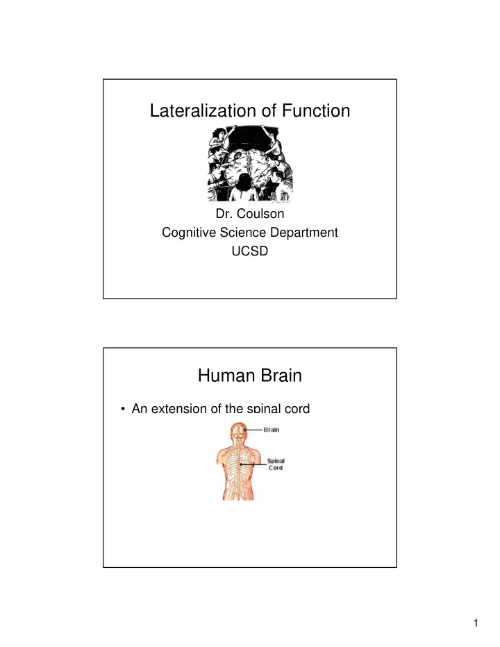
Human Brain
- An extension of the spinal cord
Lateralization of Function Dr. Coulson Cognitive Science Department - - PDF document
Lateralization of Function Dr. Coulson Cognitive Science Department UCSD Human Brain An extension of the spinal cord 1 Cerebral Hemispheres Corpus Callosum 2 Cartoon View of Brain Cerebral Lobes 3 Neurons Brain composed of
– 100 billion
– Cell body – Axon – Synapse
– Strongly vs. Weakly Lateralized
to the lower rear portion of frontal lobe, lower front portion
damage most important
to brain damage
portion of frontal lobe, adjacent to motor cortex
– Inferior frontal gyrus – Brodmann’s Areas 44/45
– 52 areas in the human brain (though some subdivided into a, b, etc)
um...'dopted...Si-sisters and mother...ball. Ball, prince um, shoe...
no, prince, yes. Cinderella hooked prince. (Laughs.) Um, um, shoes, um, twelve o'clock ball, finished.
um met.
and this. These things going in there like that. This is /sen/ things
back in this one, this one /gesh/ look at this one.
know where it is. But I don't know what's under. I know it's you couldn't say it's ... I couldn't say what it is. I couldn't say what that is. This shu-- that should be right in here. That's very bad in there. Anyway, this one here, and that, and that's it. This is the getting in here and that's the getting around here, and that, and that's it. This is getting in here and that's the getting around here, this one and
what else you'd want.
psychology.rutgers.edu/~rypma/
psychology.rutgers.edu/~rypma/
psychology.rutgers.edu/~rypma/
another stroke that resulted in paralysis of the left side of his body
– Which side did this stroke affect?
wrong with him
– Issued press release saying he hurt his left arm in a fall – Anosagnosia
medical problems from public and ran shadow government…
vantage points, a neglect patient describes different parts of the square
perspectives, but mental image lacks detail about the left half of space in each case!
Only R side active
Posner and Raichle, 1994
– Communicative disorders – Frontal damage leads to expressive disorders, trouble with grammatical complexity – Posterior damage leads to receptive disorders, trouble with meaning
– Anosagnosia – Body image disorder – Hemineglect
– Naming disruption w/stimulation in LH, not typically RH – Exact locale varies widely from individual to individual – Different languages disrupted at slightly different sites in cortex