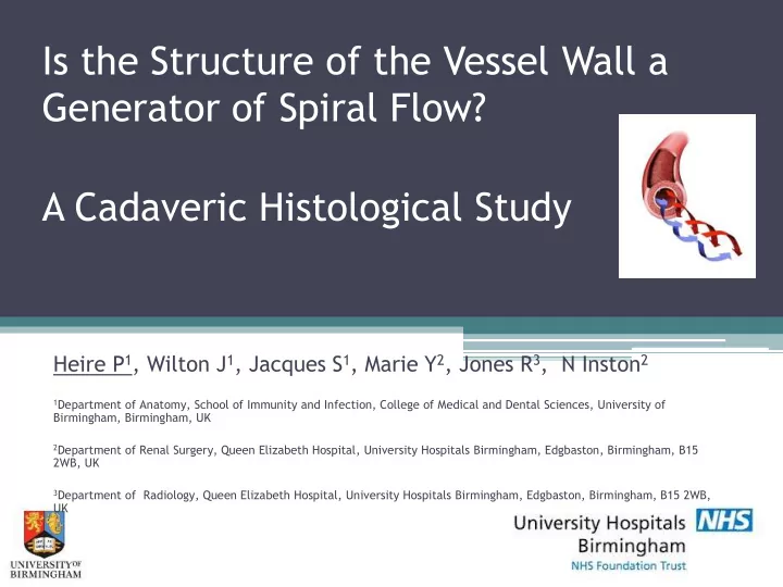

Is the Structure of the Vessel Wall a Generator of Spiral Flow? A Cadaveric Histological Study Heire P 1 , Wilton J 1 , Jacques S 1 , Marie Y 2 , Jones R 3 , N Inston 2 1 Department of Anatomy, School of Immunity and Infection, College of Medical and Dental Sciences, University of Birmingham, Birmingham, UK 2 Department of Renal Surgery, Queen Elizabeth Hospital, University Hospitals Birmingham, Edgbaston, Birmingham, B15 2WB, UK 3 Department of Radiology, Queen Elizabeth Hospital, University Hospitals Birmingham, Edgbaston, Birmingham, B15 2WB, UK
Background • Spiral laminar flow may be • In healthy individuals flow preventative against the patterns in the arterial tree development of atherosclerosis have a spiral vector (Spiral by: Laminar Flow, SLF) ▫ Reducing the laterally directed ▫ Peripheral arteries with forces on the vessel wall and angioscopy (Stonebridge and stabilising flow (Stonebridge et Brophy 1991) al 2004) ▫ Inhibiting the expression of ▫ Normal physiological finding genes involved in (Houston et al 2003, Kilner et al atherosclerosis (Chen 2002) 1993) ▫ Inhibiting the expression of adhesion molecules (Chappelle et al 1998) ▫ Loss of spiral flow associated with vascular disease (Houston et al 2004)
Background • Spiral flow is seen in the cephalic vein in brachiocephalic arteriovenous fistulas (Marie et al 2012) This implies that the vessel (artery and vein) must be capable of generating spiral flow
Aims • Assess the anatomical structure of artery and vein (muscle fibre orientation) as a potential generator of spiral flow
Methods • 4 brachial arteries and 4 cephalic veins taken from embalmed cadavers. • Sectioned in 2 angles (0˚ and 20˚) • Mounted on slides and stained using H&E • Region of the vessel tunica media chosen at random and all cell nuclei measured within that region
Methods – angular sectioning • Assessment of smooth muscle pitch • Dimensions of vascular smooth muscle cell nucleus 37µm by 2µm (Walmsley and Canham 1979) • Size of nucleus in cross- section will depend on angle at which it is sectioned. Adapted from Canham (1977)
Results Brachial artery Section angle 0˚ (n=798) 20˚ (n=1945) Median nucleus length ( μ m) 16.32 (15.67-17.00) 7.54 (7.39-7.74) (CI interval) Smooth muscle pitch = 6.04˚ to 6.28˚ Cephalic vein Section angle 0˚ (n=328) 20˚ (n=548) Median nucleus length ( μ m) 10.72 (9.76-11.92) 5.33 (5.16-5.65) (CI interval) Smooth muscle pitch = 1.37˚ to 9.33˚
Artists impression Illustration Aimee Jewitt Harris
Discussion • Conflicting evidence describing the orientation of fibres within the arterial and venous tunica media Artery: Vein: • Circumferential – Walmsley et al • Spiral – Todd et al (1983) *portal vein* (1983), Wolinsky & Glagov (1963), Arner & Uvelius (1982), Dingemans (2000), O’Connell (2008) • Spiral – Todd et al (1983), Herlihy & Murphy (1973), Borovic et al (2010), Fujiwara and Uehara (1992)
Summary • Blood vessels are capable of generating spiral flow • The alignment of muscle fibres is angular and might be the mechanism • This should be considered in design of vascular devices
Recommend
More recommend