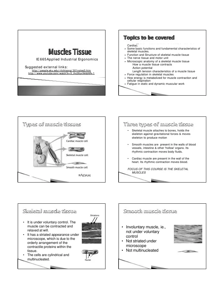

� Three types of muscle tissues: Skeletal, Smooth and Cardiac. � Some basic functions and fundamental characteristics of skeletal muscles. � Function and Structure of skeletal muscle tissue � The nerve tissue and motor unit IE 665Applied Industrial Ergonomics � Microscopic anatomy of a skeletal muscle tissue How a muscle tissue contracts Suggested external links: Action potential http:/ / people.eku.edu/ ritchisong/ 301notes3.htm Length tension characteristics of a muscle tissue http:/ / www.youtube.com/ watch? v= 0_ihc26yxN4&NR= 1 � Force regulation in skeletal muscles � How energy is metabolized for muscle contraction and cellular respiration � Fatigue in static and dynamic muscular work • Skeletal muscle attaches to bones, holds the skeleton against gravitational forces & moves skeleton to produce motion • Smooth muscles are present in the walls of blood vessels, intestine & other 'hollow' organs. Its rhythmic contraction moves body fluids. • Cardiac muscle are present in the wall of the heart. Its rhythmic contraction moves blood. FOCUS OF THIS COURSE IS THE SKELETAL MUSCLES Striations • It is under voluntary control. The muscle can be contracted and • Involuntary muscle, ie., relaxed at will. not under voluntary • It has a striated appearance under control microscope, which is due to the • Not striated under orderly arrangement of the microscope contractile proteins within the • Not multinucleated tissue. • The cells are cylindrical and multinucleated. Nuclei
• Involuntary, ie., not under • Produces motion – fundamental voluntary control characteristics of all living things • Striated appearance under microscope • Produces force (tension) • Auto-rhythmic, ie. contracts • Maintains posture – works against rhythmically without any nervous gravitational forces impulse (nerve impulse modifies • Provides joint stability the rhythm) • Produces heat as a bi- product of • Not multinucleated contraction • Rectangular in shape � Each skeletal muscle spans over one or more skeletal joints and the • Excitability - responds to stimuli (e.g., muscle contraction produces a force that tends to turn a bone nervous and other impulses) about its joint axis. � Skeletal muscles vary in size, • Contractility - able to shorten in length shape, and arrangement of fibers . They range from extremely • Extensibility - stretches when pulled tiny strands such as the stapedium muscle of the middle ear to large • Elasticity - tends to return to original masses such as the muscles of the shape & length after contraction or thigh. � A gross muscle contains skeletal extension muscle tissues, connective tissues, nerve tissues, and vascular (blood circulation) tissues. Out of these, only the muscle tissue has the contractile property. Each muscle is surrounded by a � Skeletal muscle cells (fibers), connective tissue sheath called the like other body cells, are soft epimysium. Fascia, connective tissue and fragile. The connective outside the epimysium, surrounds and tissue covering furnish separates the muscles. Portions of the epimysium project inward to divide the support and protection for the muscle into compartments. Each delicate cells and allow them compartment contains a bundle of to withstand the forces of muscle fibers. Each bundle is called a contraction. fasciculus and is surrounded by a layer of connective tissue called the � Through these tough tissues perimysium. Within the fasciculus, each contractile force of the muscle individual muscle cell, called a muscle cells are transmitted to the fiber, is surrounded by connective tissue called the endomysium. All these bone. connective tissue fuse together at the � The coverings also provide two end and forms tendon, which pathways for the passage of connects muscles to bones blood vessels and nerves .
The connective tissues, the � Skeletal muscles have an epimysium, perimysium, and endomysium extend beyond the fleshy abundant supply of blood part of the muscle to form a thick vessels, approximately 2 ropelike tendon or a broad, flat sheet- capillaries per muscle cell. like aponeurosis . Capillaries supply the The tendon form attachments from essential oxygen and muscles to the bones and aponeurosis forms connection to the connective nutrients to each muscle tissue of other muscles. fiber. Typically a muscle spans a joint � Since the capillaries spreads and is attached to bones by evenly in the muscle body tendons at both ends. One of the bones remains relatively fixed or the smaller muscles cells stable while the other end moves as a have more capillaries. result of muscle contraction. Ligaments forms joint capsules are fibrous tissues that connect bone to bone. � The nervous system is � It is the major controlling, regulatory, and composed of central communicating system in the body. If muscles are nervous system (brain power house, then the nerves are the control and spinal chord) and mechanism. peripheral nervous � It is the center of all mental activity including thought, system (containing nerve learning, and memory. � Together with the endocrine system (producing cells external to the brain hormones), the nervous system is responsible for or spinal cord). regulating and maintaining homeostasis (regulates � These, in turn, consist of internal environment so as to maintain a stable, various tissues, including constant condition) . nerve, blood, and � Through its receptors, the nervous system keeps us connective tissue. in touch with our environment, both external and internal. � Millions of sensory receptors detect changes, called stimuli, which � Axon terminals of one motor neuron innervate a number of occur inside and outside the body. They monitor such things as muscle cells that are dispersed temperature, light, and sound from the external environment. Inside randomly in the overall muscle the body, the internal environment, receptors detect variations in mass. The muscle cells and pressure, pH, carbon dioxide concentration, and the levels of the single motor neuron that various electrolytes. All of this gathered information is called innervates them make one sensory input ( afferent nervous system ). motor unit. � Sensory input is converted into electrical signals called nerve impulses that are transmitted to the brain. There the signals are � When the neuron of a motor unit sends a nerve impulse which brought together to create sensations, to produce thoughts, or to exceeds a threshold value, all the muscle cells (fibers) of the motor add to memory; Decisions are made each moment based on the unit contract together. All or none principle sensory input. This is integration. � Number of muscle cells controlled by a motor neuron varies. � Based on the sensory input and integration, the nervous system Muscles which require fine controls may have innervations of a few responds by sending signals to muscles, causing them to contract, muscle cells per motor neuron, where as, when gross force or to glands, causing them to produce secretions. production is the primary objective, motor units innvervates large � The nerve cells that send impulse to muscle cells are called motor (over hundred) number of muscles cells. nerve (efferent nervous system).
Microscopic Structure of a Muscle Cell Neucleus Sarcolema Mitochondria Contractile proteins Sarcolemma: Bi-layer lipid membrane, semi-permeable, has specialized molecules that selectively control inflow and outflow of ions � When the nerve impulse (electrical) reaches axon end, the from the extra-cellular space. permeability of the synaptic vesicle membranes at its axon Motochondria: Organelle, where ATP (Adenosine Tri-phosphate) is ends releases chemical neurotransmitter (acetylcholine). synthesized by oxidative process. ATP is only form of energy that � This chemical binds with the muscle cell membrane muscle cells can utilize to produce mechanical energy. molecules at the synaptic cleft (known as motor end plate ), and stimulates the muscle cell. Contractile proteins: Responsible for muscle contraction. T-tubules and S arcoplasmic reticulum Arrangement of Protein filaments Muscles cells are packed with myofibrils. Myofibrils are composed of two main types of myo-filaments: thick and thin. They are arranged in a very regular, precise pattern. Myosin – thick filaments Actin – thin filaments Sliding of the thin filaments over the thick filaments causes sarcomere to contract. Sarcommere: The smallest contractile unit. Models of Protein filaments Review – U-tube video � http:/ / www.youtube.com/ watch? v=EdHzKYD xrKc&feature=player_embedded
Recommend
More recommend