Introduction to Sialendoscopy and Salivary Duct Surgery Jolie - - PowerPoint PPT Presentation

introduction to sialendoscopy and salivary duct surgery
SMART_READER_LITE
LIVE PREVIEW

Introduction to Sialendoscopy and Salivary Duct Surgery Jolie - - PowerPoint PPT Presentation

11/6/2014 Disclosures None Introduction to Sialendoscopy and Salivary Duct Surgery Jolie Chang, MD Assistant Professor Department of Otolaryngology, Head and Neck Surgery University of California, San Francisco November 6, 2014


slide-1
SLIDE 1

11/6/2014 1

Department of Otolaryngology – Head and Neck Surgery

Introduction to Sialendoscopy and Salivary Duct Surgery

Jolie Chang, MD Assistant Professor Department of Otolaryngology, Head and Neck Surgery University of California, San Francisco November 6, 2014

Department of Otolaryngology – Head and Neck Surgery

Disclosures

  • None

Department of Otolaryngology – Head and Neck Surgery

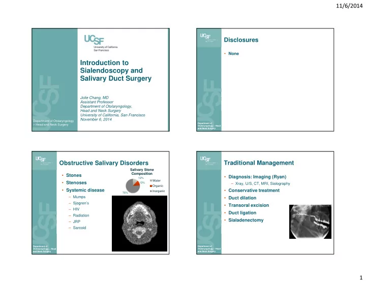
Obstructive Salivary Disorders

  • Stones
  • Stenoses
  • Systemic disease

– Mumps – Sjogren’s – HIV – Radiation – JRP – Sarcoid

12% 12% 76%

Salivary Stone Composition

Water Organic Inorganic

Department of Otolaryngology – Head and Neck Surgery

Traditional Management

  • Diagnosis: Imaging (Ryan)

– Xray, U/S, CT, MRI, Sialography

  • Conservative treatment
  • Duct dilation
  • Transoral excision
  • Duct ligation
  • Sialadenectomy
slide-2
SLIDE 2

11/6/2014 2

Department of Otolaryngology – Head and Neck Surgery

Minimally Invasive Approach

  • Endoscopic visualization of the salivary duct

– Gundlach; Konigsberger et al. HNO. 1990 – Nahlieli et al. J Oral Maxillofac Surg. 1994. – Marchal et al. NEMJ. 1999. – FDA approved 2005

Department of Otolaryngology – Head and Neck Surgery

Sialendoscopy Benefits

  • Diagnostic

– Stone location – Swelling of unknown eitology – Recurrent sialadenitis

  • Therapeutic

– Stone removal: access to posterior stones – Stenosis dilation – Spares salivary glands – Reduced risk

Department of Otolaryngology – Head and Neck Surgery

The salivary gland recovers

  • Submandibular glands removed for

sialolithiasis have normal histology. (Marchal et al 2001)

  • After stone removal, salivary function
  • improves. (Makdissi et al. 2004)
  • Animal studies: gland tissue recovers

Department of Otolaryngology – Head and Neck Surgery

Xerostomia

  • SMG saliva

– 70% of resting salivary flow – Xerostomia: Risk for dental caries

  • After SMG removal

– 22% report xerostomia in long-term follow up (Springbog and Moller. Eur Arch Otorhinolaryngol. 2013.) – Reduced unstimulated salivary flow (Cunning et al. Laryngoscope. 1998.)

slide-3
SLIDE 3

11/6/2014 3

Department of Otolaryngology – Head and Neck Surgery

Contraindication

  • Active infection

Department of Otolaryngology – Head and Neck Surgery

Clinical History

  • History
  • PMH: prior XRT, RAI, autoimmune disease
  • Exam:

– Salivary expression – Papilla location – Bimanual palpation – Mandibular tori – TMJ, trismus – Imaging

Department of Otolaryngology – Head and Neck Surgery

Sialendoscopy Setup

  • General anesthesia

– Avoid anticholinergic agents

  • Intubation

– Oral – Nasal: posterior stones in SMG

  • Preoperative Medications

– Antibiotics – Decadron

  • Positioning

ENT today. June 2008.

Department of Otolaryngology – Head and Neck Surgery

Equipment

  • Sialendoscope
  • Diagnostic sheath

– Single channel

  • Therapeutic sheath

– Working channel

slide-4
SLIDE 4

11/6/2014 4

Department of Otolaryngology – Head and Neck Surgery

Equipment (Walvekar)

Department of Otolaryngology – Head and Neck Surgery

Technique – the Papilla

  • Identify the papilla

– Gland massage – Symmetry – Magnification – Inject papilla – Methylene blue

  • Papilla dilation

– Start with smallest dilator 0000 up to size 3-4 – Avoid making pseudo

  • penings with

needle/forceps

Department of Otolaryngology – Head and Neck Surgery

Technique - Sialendoscopy

  • Setup

– Orient camera – Brightness

  • Introduce sialendoscope

– Saline irrigation – no air – Small movements – Backing out helps to find lumen – Advance under visualization – Be careful of teeth, mandibular tori

Department of Otolaryngology – Head and Neck Surgery

Examination

  • Ducts and branches
  • Stones

– Location – Mobility – Number

  • Stenoses

– Location – Extent

  • Other

– Foreign body – Lesions

slide-5
SLIDE 5

11/6/2014 5

Department of Otolaryngology – Head and Neck Surgery Department of Otolaryngology – Head and Neck Surgery

Stones – Tools for Management

  • Basket/ Dochotomy
  • Lithotripsy: Laser

– Forceps – Endoscopic drill

  • Combined approach

Department of Otolaryngology – Head and Neck Surgery

Lithotripsy

  • Impacted large stones
  • Fragmentation:

– Forceps; Hand Drill – Laser Ho:YAG – ECSWL – Endoscopic Intracorporeal SL

  • Lithotripsy

– Slow – Can damage duct and scope – Multiple sessions required

Department of Otolaryngology – Head and Neck Surgery

Combined approach

  • Due to size & location, stone may not be

amenable to endoscopic management

  • Sialendoscopy to localize and characterize the

stone

  • Submandibular

– Posterior sialodochtomy over palpable stone

  • Parotid

– Transoral sialodochotomy to release distal stones – Transfacial approach for impacted proximal stones

slide-6
SLIDE 6

11/6/2014 6

Department of Otolaryngology – Head and Neck Surgery

Combined Approach

Marchal F. Laryngoscope 2007.

Department of Otolaryngology – Head and Neck Surgery Department of Otolaryngology – Head and Neck Surgery

Salivary Duct Stenosis

  • Koch M et al. Oto HNS 2005

– Sialendoscopy in 103 cases with negative imaging (U/S) – 33% with stones – 56% with stenoses

  • Causes of stenosis

– Iatrogenic: prior surgery – Inflammatory: autoimmune, XRT, RAI

Marchal et al. J Stoma 2011.

Department of Otolaryngology – Head and Neck Surgery

Stenosis Management

  • Diagnosis

– Sialography – Sialendoscopy: determine location, extent, length

  • Treatment: Dilation

– Dilation: Hydraulic, balloon, – Stents, steroids – Sialodochotomy – Combined Approach: Excision, Vein graft – Sialadenectomy

slide-7
SLIDE 7

11/6/2014 7

Department of Otolaryngology – Head and Neck Surgery

Conclusions - Sialendoscopy

  • Minimally invasive, gland sparing approach
  • Diagnostic and therapeutic
  • Treatment of sialolithiasis and stenosis
  • Reduced need for sialadenectomy