
SLIDE 1
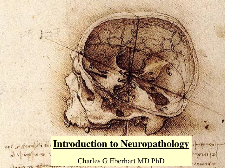
Introduction to Neuropathology
Charles G Eberhart MD PhD
Introduction to Neuropathology Charles G Eberhart MD PhD OUTLINE - - PowerPoint PPT Presentation
Introduction to Neuropathology Charles G Eberhart MD PhD OUTLINE Cellular components of the CNS Pathology of Neurons Pathology of Glia Microscopic appearance of common CNS disease processes Introduction to CNS development
Charles G Eberhart MD PhD
Neurons Drawn by Franz Nissl 1860-1919
and prominent nucleolus (“fried egg”)
(nuclei, ganglia) or in layers
Dendritic Tree
Cerebellar granule neurons
Cerebellar Purkinje Neurons Cerebral Cortical Neuron
and insulate axons.
nuclei with dense chromatin
Oligos
and grey matter
vessels, the pia and ependyma (glia limitans)
suppliers of nutrients, and physical barriers
to oval and slightly larger than those of oligodendrocytes
Astrocyte Neuropil = “nerve felt”
Andreas Vesalius 1514-1564
lining the ventricular system
and CSF
transport between the brain and CSF Ependymal Cells
act as a fixed macrophage/ monocyte system in the brain
response to infection/injury
cells
Resting Activated Phagocytic Neuronophagia
the ependyma that secrete CSF
epithelium covering vascular cores
CSF barrier
per hour
the subarachnoid space
Skull Dura Subarachnoid Space Pia Arachnoid
inner skull periostium
up of meningothelial cells and connective tissue
drapes over the brain
the entire cortical surface, and invests arteries as they penetrate the brain
the arachnoid and pia
Cortical Surface Arachnoid Artery Subarachnoid Space Pia CSF flows out of the sub- arachnoid space into the dural sinuses through the arachnoid granulations protruding into the sinuses Arachnoid Granulation Arachnoid cap cells attached to the sinus endothelium
Fixation of CNS tissue in potassium bichromate with application of silver nitrate
Santiago Ramon y Cajal (1852-1934), Barcelona and Madrid, Spain
Ramon y Cajal improved on Golgi’s silver stain, and developed a gold chloride-mercury stain for astrocytes
Golgi and Ramon y Cajal shared the 1906 Nobel Prize for Medicine In recognition of their Work on the structure Of the nervous system
Synaptophysin GFAP (Neuronal) (Glial)
Glia - GFAP (Glial Fibrillary Acidic Protein) Neurons - Synaptophysin, NeuN Proliferation – Ki67 (MIB-1) Microglia/Macrophages – CD68 (KP1), HAM56 Lymphoid Cells – CLA, CD3 (T Cells), CD20 (B Cells) Infectious Agents – Toxoplasma, Adenovirus, JC Virus Inclusion Bodies – Ubiquitin, α-synuclein, Tau
Immunohistochemical Stains Other Stains Myelin – Luxol Fast Blue Alzheimer Dz - Hirano Silver Fungi – Methenamine silver (GMS)
MIB-1 GFAP
Johns Hopkins Department of Pathology Patient: John Doe Procedure Date: 1/1/2002 Part 1-3: Temporal Mass (Biopsy): Frozen Section Diagnosis: Low grade neoplasm Final Diagnosis: Ganglioglioma, WHO Grade I, See Comment Comment: The tumor has a solid, non-infiltrating architecture, with no intra-tumoral axons detected using SM31 immunostains. Atypical neuronal and glial cells are present in the lesion, as evidenced by positive synaptophysin and GFAP immunostains. The MIB-1 proliferation index is low (1-2%)
Apoptosis in Neuroblastic Tumor Fragmented Chromatin in Dorsal Root Ganglion Neuron
into “apoptotic bodies”
Heat, Toxic Agents, Hypoglycemia, Hypoxic/Ischemic Damage Hippocampal Ischemia Red Neurons
(hippocampus), Cortical layers 3 & 5, and Purkinje Cells are especially vulnerable
within approximately 12 Hours
also die, and the necrotic region is cleared away by macrophages
(Injury Induced Cell Death)
LFB CD68
A LFB myelin stain and CD68 macrophage immunostain highlight the axonal degeneration in the crossed and uncrossed corticospinal tracts in Amyotropic Lateral Sclerosis (ALS)
Huntington’s Dz Parkinson’s Dz Lewy Body Ubiquitin + Inclusion
Cytoplasmic
Nuclear
Reactive Astrocytosis A non-specific reaction to infection, seizures, autoimmune disease, infarction, etc Fibrillary Gliosis Proliferation of reactive astrocytes Piloid Gliosis Seen around spinal cord cavities (syrinx) And other long-standing reactive gliosis In cerebellum and hypothalamus. Also In Alexander’s disease Reactive Astrocytosis Piloid Gliosis
JC Virus Immunostain Infected Oligodendrocytes
This last section in intended to introduce you to the microscopic appearance of several common CNS diseases. More detailed examples and explanations will be provided in later lectures.
Hours – Days: Neurons become eosinophilic and shrunken Neutrophils infiltrate the lesion Days - Weeks: Neurons gone, macrophages infiltrate lesion Reactive astrocytosis around edge Weeks – Months: Cystic cavity Macrophages Old Cystic Infarction
Meningitis Abcess
Perivascular Lymphocytes Microglial Nodule
echo, coxsackie, herpes, mumps, measles adenovirus, polio, VZV, EBV, CMV, rabies, arboviruses, JC, HIV
intraparenchymal lymphocytes
and microglial nodules also commonly present Herpes
Macrophages Loss of myelin on HE/LFB stain
pallor on LFB stain
have sharp borders
reactive astrocytes found in plaque
All of the cell types in the brain Can give rise to tumors
Glial tumors are the most common Malignant lesions Oligodendroglioma Astrocytoma
It has long been thought that brain tumors resemble (and perhaps arise from) stem/precursor cells
A Classification of the Tumors of the Glioma Group on a Histogenetic Basis with a Correlated Study of Prognosis. (1926)
Harvey Cushing
Cerebellar Anlage VZ
Conventions & Terminology
R L
As If From Front As If From Front
R L L R
As If From Back Diagram MRI-CT-Radiograph Pathology Specimen
Conventions & Terminology
R L
MRI As If From Bottom Of Feet
L R
OD OS Visual Field Pathways As If From Top Of Head
IMAGI GING NG: MR MRI B I Brain wi with in intrapa parenc enchy hym al al hem hemorrhage from m mycotic ic aneurys ysm Elucida lucidate tes… 1) 1) pathoa thoana natom tomy, 2) 2) pathol thology
3) 3) pathoph thophysiol
4) 4) clinical ri risk CLOT MIDL MIDLINE SH SHIFT EDEM EMA
Non contrast or plain imaging appearances
Scan Uses CSF Lesion Blood Bone
CT Rapid screen Dark Dark White White T1 MRI Anatomy Dark Dark White Dark T2 MRI Lesion ident. White White Varies with age
Dark FLAIR Lesion ident. Dark White Varies with age
Dark
– Major mass effect with midline shift – (Obstructive) hydrocephalus
– E.g., Subarachnoid, Intraparenchymal
– Skull fractures – Bone erosion from infection
– Bullets and other metal
acutely
– CT Angiography (e.g. for acute stroke)
patient cannot get an MRI
– Pacemaker or other paramagnetic retained foreign body – Severe claustrophobia – None available
CT, Noncontrast (soft tissue w indow ) CT: skull is w hite Soft Tissue Window : skull detail not visible, CSF black, gray vs. w hite matter discrimination fuzzy Noncontrast: vessels are inapparent
NLM Visible Human Project
CT, Noncontrast (bone w indow ) CT: skull is w hite Bone Window : skull detail visible, soft tissues indiscernable (CSF, gray & w hite matter, vessels)
CT Subarachnoid Hemorrhage Normal CT
Bright BLOOD in the peri-mesencephalic SA spaces
Dilated temporal horn lateral ventricle
Normal CT Bone Window s @
Note that MRI is useless for imaging bone rel. to CT
Normal MRI T1 contrast @
pathology
– including lesions without large mass effect (esp. white matter disease)
lesions
(Diffusion Weighted Imaging [DWI])
(MR angiogram
[MRA/V])
MRI, T1, noncontrast MRI: skull is black (scalp fat & bone marrow w hite) TI: CSF black, & differentiation of gray vs. w hite matter good Noncontrast: vessels inapparent
MRI, T1, Contrast MRI: skull is black (scalp fat & bone marrow w hite) TI: CSF black, & differentiation of gray vs. w hite matter good; gray matter darker than w hite matter Contrast: vessels w hite
T1 Noncontrast T1 Contrast
Note subtle appearance of contrast in blood vessels
MRI, T2, noncontrast MRI: skull is black (scalp fat & bone marrow w hite) T2: CSF w hite, & differentiation of gray vs. w hite matter fair; w hite matter darker than gray matter Non Contrast: vessels inapparent (small black flow voids)
MRI FLAIR (fluid attenuated inversion recovery) MRI: skull is black (scalp fat & bone marrow w hite) FLAIR: CSF black, & differentiation of gray vs. w hite matter poor; w hite matter darker than gray matter
marrow fat in diploic space Essentially a T2 image w ith the CSF ‘averaged’ out
MRI scan: T2 vs. FLAIR – Why use FLAIR?
Note that pathology ‘stands out’ w hen CSF is ‘averaged out’ of the image As a result, FLAIR images end up being more sensitive, but less specific