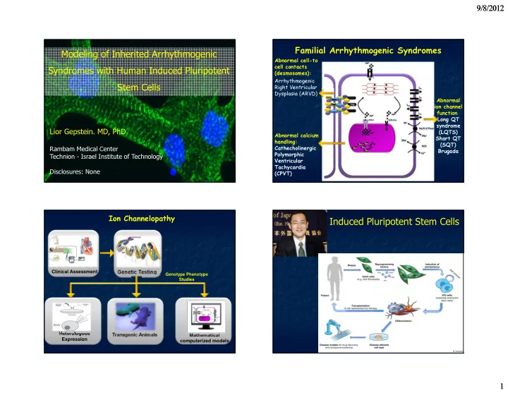
induced pluripotent stem cells
play

Induced Pluripotent Stem Cells Clinical Assessment Genetic Testing - PowerPoint PPT Presentation

9/8/2012 Familial Arrhythmogenic Syndromes Modeling of Inherited Arrhythmogenic Abnormal cell-to cell contacts Syndromes with Human Induced Pluripotent (desmosomes): Arrhythmogenic Stem Cells Right Ventricular Dysplasia (ARVD) Abnormal


  1. 9/8/2012 Familial Arrhythmogenic Syndromes Modeling of Inherited Arrhythmogenic Abnormal cell-to cell contacts Syndromes with Human Induced Pluripotent (desmosomes): Arrhythmogenic Stem Cells Right Ventricular Dysplasia (ARVD) Abnormal ion channel function: Long QT syndrome Lior Gepstein. MD, PhD (LQTS) Abnormal calcium Short QT handling: (SQT) Cathecholinergic Rambam Medical Center Brugada Polymorphic Technion - Israel Institute of Technology Ventricular Tachycardia Disclosures: None (CPVT) Ion Channelopathy Induced Pluripotent Stem Cells Clinical Assessment Genetic Testing Genotype Phenotype Studies Heterologous Transgenic Animals Mathematical Expression computerized models 1

  2. 9/8/2012 Cardiomyocyte Differentiation of hiPSCs hIPSCs-Derived Cardiomyocytes Tra-I-60 hiPSCs-colonies Fibroblasts α -actinin cTnI 30 mV hiPSCs-derived cardiomyocytes 1 sec * Zwi et al., Circulation 2009 Zwi et al., Circulation 2009 Reprogramming Calcium Handling in hiPSCs-CMs Somatic Cells iPS Cells Sarcomeric α-actinin Caffeine puff RyR2 Patient/Disease Specific Structural analysis Patch-clamp Cardiomyocytes 250 ms Multielectrode array Gene expression analysis recordings 1 F/F 0 Confocal Calcium Imaging 5 sec Itzhaki, et al. PLoS One 2011 2

  3. 9/8/2012 Long QT Syndrome LQTS-Fibroblasts PVC QTc-520ms * C T (Ala 614 Val) * KCNH2 gene Itzhaki I, et al. Nature 2011 1500 Ventricular * Action Potential Recordings from Multielectrode Array (MEA) Recordings * APD (msec) Control and LQTS hiPSCs-CMs * 1000 Spontaneous APs 500 Control Control LQTS Ventricular Atrial Nodal Ventricular Atrial Nodal 0 APD 50 APD 70 APD 90 0 mV 1000 Atrial * Control 0 mV 800 * APD (mseC) LQTS 600 * 30 mV 400 200 Control 400 ms LQTS 0 APD 50 APD 70 APD 90 800 Nodal LQTS 0 mV 30 mV 600 APD (msec) 400 400 ms Control 200 FPD- 0.31 sec Control LQTS 0 Ventricular- Paced at 0.5Hz APD 50 APD 70 APD 90 LQTS Control LQTS FPD- 1.01 sec 1500 Ventricular - Paced at 0.5 Hz * * APD (msec) 1000 * 500 30 mV Control The observed prolongation (in APD and cFPD) is the LQTS 0 characteristic electrophysiological signature of the LQTS. 1 sec APD 50 APD 70 APD 90 3

  4. 9/8/2012 Α614ς Early After Depolarizations (EADs) in the LQTS Control Ventricular Atrial Nodal 0 mV LQTS 30 mV 400 ms LQTS Ventricular EAD Atrial EAD 0 mV 30 mV 400 ms Itzhaki I, Meizels L, Huber I, et al. Nature 2011 Itzhaki I, Meizels L, Huber I, et al. Nature 2011 Pinacidil (K ATP Channel Opener) Ameliorating Effects on the LQTS-hiPSCs-CMs Development of Triggered-Activity in the LQTS hiPSCs-CMs LQTS – Baseline LQTS – Pinacidil Triggered beat Triggered beat EAD EAD 30 mV 20 mV 1 sec LQTS – Baseline 5 sec LQTS – Pinacidil 20 mV 5 sec 2 sec 2 sec Itzhaki I, Meizels L, Huber I, et al. Nature 2011 Itzhaki I, et al. Nature 2011 4

  5. 9/8/2012 Catecholaminergic Polymorphic Ventricular Tachycardia (CPVT) * * T G (Met 4109 Arg) (M4109R) heterozygous CPVT- missense mutation Type I AP OCT4 NANOG TRA1 –60 SSEA 4 CPVT- Type II Liu, et al. J Mol Cell Cardiol, 2009 Itzhaki I, Meizels L, et al. J Am Coll Cardiol (accepted) Undiff CMs CPVT hiPSCs-CMs are Arrhythmogenic cTnI CPVT OCT4 CPVT hiPSC-CM CPVT hiPSC-CM DADs (phase 4) Late phase 3 ADs % of cells presenting DADs NANOG NKX2-5 80% 70% * MLC2V RyR α - actinin merged 60% Control α - actinin 30 mV 50% 30 mV MYH6 40% 1 sec 1 sec 30% MYH7 20% Control hiPSC-CM Control hiPSC-CM DADs (phase 4) CTNI 10% 0% CPVT Control β-ACTIN RyR α - actinin merged Ventricular Atrial Nodal 30 mV 30 mV 1 sec 1 sec CPVT 0 mV 30 mV Control - Paced CPVT- Paced 500 ms Control 0 mV 30 mV 30 mV 30 mV 1 sec 1 sec 500 ms Itzhaki I, Meizels L, et al. J Am Coll Cardiol (accepted) Itzhaki I, et al. J Am Coll Cardiol (accepted) 5

  6. 9/8/2012 Adrenergic Stimulation Enhances Arrhythmogenic Role of intracellular calcium stores potential of the CPVT hiPSCs-CMs CPVT hiPSC-CM CPVT hiPSC-CM Forskolin Baseline TA Baseline Thapsigargin 5 mV 30 mV 1 sec 1 sec TA Drug testing using CPVT-hiPSCs-CMs: Flecainide 30mV 2 sec CPVT hiPSC-CM CPVT hiPSC-CM Isoproterenol Isoproterenol Baseline Flecainide Baseline TA 30mV 30 mV 30 mV 30mV 1 sec 1 sec 1 sec 2 sec Itzhaki I, Meizels L, et al. J Am Coll Cardiol (accepted) Laser Confocal Calcium Imaging Calcium Imaging of the CPVT- hiPSCs-CMs – Adrenergic Stimulation CPVT-hiPSCs-CMs Control-hiPSCs-CMs CPVT-hiPSCs-CMs CPVT-hiPSCs-CMs CPVT-hiPSCs-CMs 5 F/F0 1 F/F0 Isoproterenol Propanolol Baseline 2 sec 2 sec 2 F/F0 2 F/F0 2 F/F0 1 F/F0 1 F/F0 2 sec 2 sec 2 sec 8 sec 4 sec 1 F/Fo 8 sec Itzhaki I, Meizels L, et al. J Am Coll Cardiol (accepted) Itzhaki I, Meizels L, et al. J Am Coll Cardiol (accepted) 6

  7. 9/8/2012 Store Overload Induced Calcium Release (SOICR) Store Overload Induced Calcium Release (SOICR) CPVT hiPSC-CM Control hiPSC-CM 0.1mM 2 F/F0 2 F/F0 2 F/F0 2 F/F0 0.2mM + 2 F/F0 2 F/F0 0.3mM 2 F/F0 2 F/F0 0.5mM 2 F/F0 2 F/F0 1 mM 2 F/F0 2 F/F0 2 mM 2 F/F0 2 F/F0 3 mM 2 F/F0 2 F/F0 4 mM 2 sec 2 sec Priori SG, Chen SR, Circ Res. 2011 Itzhaki I, Meizels L, et al. J Am Coll Cardiol (accepted) Store Overload Induced Calcium Release (SOICR) % of cells presenting Ca 2+ transients 120% Control CPVT 100% * * 80% * 60% * 40% * * 20% 0% 0.1 0.2 0.3 0.5 1 2 3 4 Bath Ca 2+ concentration (mM) Itzhaki I, Meizels L, et al. J Am Coll Cardiol (accepted) 7

  8. 9/8/2012 CPVT-2 hiPSCs-CMs are Arrhythmogenic Laser Confocal Calcium Imaging of CPVT-2 hiPSCs-CMs CPVT2 hiPSC-CM Baseline 5 Φ/Φ0 5 Φ/Φ0 CPVT2 hiPSC-CM Post-pacing 8 σεχ 4 σεχ CPVT2 hiPSC-CM 0.1mM Ca 2+ Φ/Φ0 CPVT2 hiPSC-CM 2 Isoproterenol 120% Control Φ/Φ0 0.2mM Ca 2+ % cells presenting Ca2+ transients 2 100% CPVT2 Φ/Φ0 0.5mM Ca 2+ 2 80% Φ/Φ0 2 1mM Ca 2+ 60% Φ/Φ0 2 2mM Ca 2+ 40% Φ/Φ0 20% 3mM Ca 2+ 2 CPVT2 hiPSC-CM Φ/Φ0 0% Isoproterenol + propanolol 4mM Ca 2+ 2 0.1 0.2 0.5 1 2 3 4 2 sec Bath Ca2+ concentration (mM) Modeling of ARVC with hiPSCs Modeling of ARVC with hiPSCs ARVC ARVC hiPSCs Cardiomyocytes PKP-2 14% PKP2/cTNI Signal Intensity 12% 10% PKP-2 PKP-2 8% 6% 4% v 2% 0% Control ARVC Insertion Stop mutation codon Plakoglobin 7% Plakoglobin Plakoglobin PKG/cTNI Signal Intensity 6% 5% c.972InsT 4% A324fs335X/N 3% 2% 1% 0% Control ARVC Control ARVC 8

  9. 9/8/2012 Modeling of ARVC with hiPSCs Modeling of ARVC with hiPSCs Desmosomal gap width 40 * 30 nm 20 10 ARVC ARVC ARVC ARVC hiPSCs Cardiomyocytes hiPSCs Cardiomyocytes 0 Control ARVC Total desmosome width D ** 200 M 150 S nm D S D 100 D 50 200nm 200nm 0 Control ARVC Control ARVC Modeling of ARVC with hiPSCs Summary � The disease phenotypes presented by the LQTS, CPVT, ARVC, and Pompe Disease patients Exposure to Lipogenic stimuli at bedside could be recapitulated in-vitro using ARVC ARVC the hiPSC approach hiPSCs Cardiomyocytes � LQTS: APD prolongation, EADs, and triggered activity PPARG � CPVT: Abnormal Ca leak, DADs, triggered activity 500 � ARVC: Abnormal desmosomes 450 ∗ 400 350 RQ 300 250 200 150 100 50 0 Control ARVC 9

  10. 9/8/2012 Summary Summary � The hiPSC approach may bring a unique value to the � The hiPSC-CM model can be used to confirm or field of drug development and personalized medicine; provide new insights into disease mechanisms screening the effects of potential disease � LQTS: Confirm the role of EADs and TA aggravators and existing and novel therapies in a � CPVT: Role of DADs and TA, role of the RyR2 patient-specific manner mutation in altering the threshold for SOICR � ARVC: Correlation between the degree of desmosomal abnormalities and lipid accumulation. Potential role for adipogenesis in the disease process Tissue Engineering: Zimmermann et al. Limitations and challenges of iPSC technology (Circ. Res, 2002) • Incomplete reprogramming & Epigenetic memory • Cardiomyocyte Heterogeneity • Early-stage cardiac phenotype • Need for a more clinically- realistic three-dimensional multicellular highly- structured engineered cardiac tissue 10

  11. 9/8/2012 Humane Engineered Myocardium Tiburcy et al. (unpublished) Joseph Itskovitz-Eldor Izhak Kehat Michal Amit Oren Caspi Jackie Schiller Leonid Kheimovitz Rafael Beyar UCSF – Jeffery Olgin Ilanit Izhaki Haim Hammerman Emily Wilson Manhal Habib Monther Bolous Chunua Ding Irit Huber Hana Mandel Amira Gepstein Gil Arbel Aya Lange Wolfram Zimmermann Izhak Mizrahi Thomas Hescenhagen Limor Zvi Leonid Meisels Jonathan Satin Oren Feldman 11

  12. 9/8/2012 Thank You 12

Download Presentation
Download Policy: The content available on the website is offered to you 'AS IS' for your personal information and use only. It cannot be commercialized, licensed, or distributed on other websites without prior consent from the author. To download a presentation, simply click this link. If you encounter any difficulties during the download process, it's possible that the publisher has removed the file from their server.

Recommend


More recommend