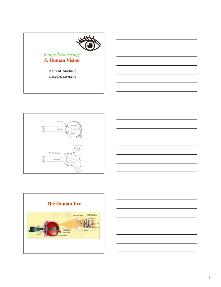

Image Processing : 5. Human Vision Aleix M. Martinez aleix@ece.osu.edu The Human Eye 1
2
Diopters 3
Simple and complex cells TT]E EYE Stimulus: on Stimulus: on The more of a given region, on or off , the stimulus filled, the greater was Le-li: Four recordings fronr a typical on- the response, so that maximal on responses center retinal ganglion cell. Each record is a were obtained to just the right size single circular spot, and maximal off responses sweep of the oscilloscope, whose to a ring ofjust the right dimensions duration is 2.5 seconds. For a sweep this (inner and outer diameters). Typical recordings of responscs to such stimuli are slow, the rising and falling phases of the shown on this page. The center and surround regions interacted in an anrago- impulse coalesce so that each spike appears nistic way: the effect of a spot in the center was diminished by shining a second as a vertical line. To the left the stimuli are spot in the surround-as if you were telling the cell to fire faster and slower ar shown. In the resting srate at thc top, there is no stimulus: firing is slow and morc or the same time. The most impressive demonstration of this interaction between less random. The lower three records show center and surround occurred when a large spot covered the entire receptive responses to a srnall (optin-rum size) spot, a ficld of the eanglion cell. This evoked a response that u,as much weaker rhan larple spot covering the receptive-field cen- the rcsponse to a spot just filling the center; indeed, for some cells the effects of ter and surround, and a ring covering the stimulating the two regions cancelled cach other cornpletely. surround only. Rigftr: Responses of an off- center retinal ganglion cell to the sante set An of-tenter cell had just the opposite behavior. Its receptive field consisted of stinruli shown at the left. of a small ccnter frorn which off responses were obtained, and a surround that gave on responscs. The two kinds of cells were intermixed and seemed to be equally common. An off-center cell discharges at its highest rate in response to a black spot on a white background, because we are now illuminating only the surround of its receptive field. In nature, dark objects are probably jusr as common as light ones, which may help explain why information from the retina is in the form of both oll-center cells and off-center cells. lf you make a spot progressively larger, the rcsponse inrproves until the receptive-field center is filled, then it starts to decline as more and more of the surround is included, as you can see from the graph on the ltcxt page. With a spot covering the etrtire field, the center either just barely wins oLlt over the surround, or the result is a draw. This effect explains why neurophysiologists CHAI)T'Ell I Optic nerve Optic chiasm Optic tract Lateral geniculate nucleus Optic radiations Prirnary visual cortex 7 138 CHAPTER The corpus callosum is a thick, bent plate of axons near the center of this brain sec- tion, made by cutting apart the human ce- rebral hemispheres and looking at the cut surface. Occasionally it may be com- condition called agenesis of the corpus callosum. either to treat epilepsy (thus pre- pletely or parrially cut by the neurosurgeon, that begin in one hemisphere from spreading to venting epileptic discharges the other) or to make it possible to reach a very deep tumor, such as one in the had neurologists and psy- pituitary gland, from above. In none of these cases had even suggested (perhaps not seri- chiatrists found any deficiency; someone function of the corpus callosum was to hold the two cere- ously) that the sole bral hemispheres together. Until the r95os we knew little about the detailed the two cerebral connections of the corpus callosum. It clearly connected crude neurophysiology it was thought hemispheres, and on the basis of rather join precisely cortical areas on the two sides. Even cells in the to corresponding striate cortex were assumed to send axons into the corpus callosum to termi- on the opposite side. nate in the exactly corresponding part of the striate cortex student studying under psychologist In 1955 Ronald Myers, a graduate at the University of Chicago, did the first experiment that re- Roger Sperry in a box vealed a function for this immense bundle of fibers. Myers trained cats 4
THE CORPUS CALLOSUM AND STET{EOPSIS Here the brain is seen from above. On the right side an inch or so of the top has been lopped off. We can see the band of the cor- pus callosum fanning out after crossing, and joining every part of the two hemi- spheres. (The front of the brain is at the top of the picture.) containing two side-by-side screcns onto which hc could project imagcs, for example a circle onto onc screcn and a square onto thc other. He taught a cat to press its nose against the screen with the circle, in prefcrcnce to the one with by rewarding correct responses with food and punishing mistakes the square, mildly by sounding an unpleasantly loud buzzer and pulling the cat back from the screen gently but firmly. By this method the cat could be brought to a fairly consistent performance in a few thousand trials. (Cats learn slowly; a THE CORPUS CALLOSUM AND STEREOPSIS In his experiment, Whitteridge cut the right optic tract. For information to get from either eye to the right visual cortex, it now has to go to the left visual cortex and cross in the corpus callosum. Cooling either of these areas blocks the flow of nerve impulses. Corpus callosum Cortex side world-except, of course, for any input that area might receive from the left occipital lobe via the corpus callosum, as you can see from the illustration on this page. He then looked for responses by shining light in the eyes and recording from the right hemisphere with wire electrodes placed on the corti- cal surface. He did record responses, but the electrical waves he observed appeared only at the inner border of area 17, a region that gets its visual input from a long, narrow, vertical strip bisecting the visual field: when he used smaller spots of light, they produced responses only when they were flashed in The Arnolfini Portrait Jan van Eyck, 1434 5
74 Perception and Artistic Style The t as vi Two both , \ , 2 ' t l \ world t,. \ \ ./r/ \ ,lr' . horizc '\ 1 \ / / ' ' \ \. ./i' /,/' ./ € vok € r . \ L / . t . . " \\ \ /.' / t ' , t horizc -Y.r' \ \ t , identir that or lines i 2.6). t angle paper-r constn be four Figure 4.3 The diagram shows the inconsistencies in perspective in the van Eyck porhait shown in figure 1..1. one would expect them to be readily noticeable but in fact they are not Figure 4 illusion. La Crau Vincent van Gogh Cues Intrinsic to the Eye Perception and Artistic Style 1,39 i i ---k-:-] .i*.* * ,/ll " ffi r-4#'6i, '(),i0ffi, 1, "Le Crau". (Reproduced by Amsterdam). Figure 7.38 Focal points implicit in van Gogh's Le Crau are shown in this diagram. 6
7
Recommend
More recommend