
SLIDE 1
I.HISTORY ULTRASOUND SCANNING TECHNIQUE for EARLY PREGNANCY - - PowerPoint PPT Presentation
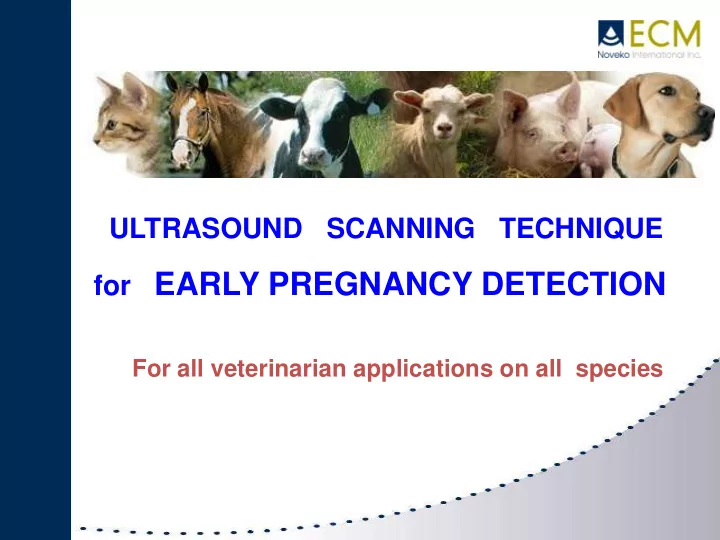
I.HISTORY ULTRASOUND SCANNING TECHNIQUE for EARLY PREGNANCY DETECTION For all veterinarian applications on all species BOVINE EQUINE MARKET Practice for bovine ultrasound scanning is essentialy made with linear rectal probes, but it is
palpation.
can take propriate measures to get cows bred more quickly.
56 days) cows often retain their corpus luteum and therefore delays return to estrus (luteolytic drugs allowing expel
1.000 cows farm pays for a new ultrasound system with the savings on feed cost after 2 months.
millions US dollars/year in the US dairy industry !!!
which is opn past 100 days in milk.
the uterus and therfore risk of inducing embryonic mortality.
Early pregnancy diagnosis: 30 days and less with experience Twin pregnancy Fœtal sexing from 55 to 90 days Non-pregnancy diagnosis Ovary and corpus luteum examination Cyst, Follicles control Metritis Piometria Early embryonic death… Back fat measurement (At the level of the back triangle) Bladder diagnosis Teats lesion control Genital bull tract diagnosis Umbilical cord control on calves
With linear rectal probe
With sector rectal probe
Pregnancy 22 days
Pregnancy 26 days Pregnancy 27 days Only 6mm length
Pregnancy 34 days Pregnancy 32 days - about 1cm
Pregnancy 40 days Twins pregnancy – 38d 2 foetus
Pro oestrus 19 days Metaestrus 36 hours
Embryonic Death Metritis
Fœtal sexing
Male 67 days Female 57 days scrotum Future lips
Teat Lesions control
Bull Tracts Exam
(Testicle Ipoplasia)
Emaciated Body condition scoring (BCS) Back fat (mm) Very Poor 1.0 < 5 Poor 1.5 5 2 10 Moderate 2.5 15 Good 3 20 Very Good 3.5 25 Fat 4 30 Adipose 4.5 35 Obese 5 > 35
65 days 85 days
Empty horns Cotyledons – 4 months
Reduce number of empty cows Confirm pregnant cows from 30 days Reduce gap between 2 calvings Manage dry-off period Reduce feeding expenses Know calving dates Make surveillance of heats easier and handle reproduction better Scrap empty cows at the right time Determine fœtal gender Check twin pregnancies and pay more attention during calving Sell animals guarantied pregnant Act rapidly on cows with problems (cyst – metritis – embrionic death…) Date pregnancy and make calving batches
Ultrasound scanning on ovine and caprine was essentially made with linear abdominal probes A convex probe of 3.5 MHz that allows a diagnosis from 15 to 18 cm depth is the most adapted However, it is possible to make pregnancy controls with sector probes (IMAGO.S)
Pregnancy diagnosis Non pregnancy diagnosis (Cyst, follicle, Metritis) Detection of twins, triplets Detection of pseudo pregnancies (on goats) Back fat measurement Early pregnancy diagnosis:
Determination of pregnancy stage Bladder diagnosis Multiple (fetal counting):between 40 and 80 days Back fat measurement
Pregnancy 35 days Twins 60 days
Pregnancy 75 days Pseudo pregnancy
Ultrasound scanning is used for long time and not only for reproduction purpose,but also for othopedics (tendons) trouble and even abdominal diagnosis
CLIP HAIRS USE ALCOHOL And GEL PAD
11 days pregnancy 26 days
REPRODUCTION EXAMS TENDONS DIAGNOSIS ABDOMINAL EXAMS
The use of ultrasound scanner is not used a lot for pregnancy control and detect the number of puppies.The main applications are abdominal controls and more and more about cardiology aspects
KIDNEY CAT HEAD
ASCITIS (fluid in abdomen)
liver
DUEDONUM
SPLEEN
KIDNEY STOMACH
Liqui
256 tones of grey
Better penetration Higher resolution
Ultrasound scanning is of a great interest in pregnancy control for bovine and equine gynecology,also camels and small ruminants. It’s a great help in herd management , it allows to make early pregnancy
The technician (Veterinarian, breeder, or service provider) can be precise in his diagnosis and adapt his treatment to the problem He will then be more efficient, and if this is a vet or a service provider) he will be able to advise his customer better ECM’s goal is to provide the best answer when it’s about ultrasound scanning. There is always a portable system and one or several probes adapted to your request. THANKS