I have no relevant relationships Diagnosis and Management - - PowerPoint PPT Presentation

i have no relevant relationships
SMART_READER_LITE
LIVE PREVIEW

I have no relevant relationships Diagnosis and Management - - PowerPoint PPT Presentation

5/26/2017 Gastric Polyps: I have no relevant relationships Diagnosis and Management requiring disclosure Gregory Y. Lauwers, MD Senior Member H. Lee Moffitt Cancer Center & Research Institute Tampa, FL


slide-1
SLIDE 1

5/26/2017 1

Gastric Polyps:

Diagnosis and Management

Gregory Y. Lauwers, MD Senior Member

  • H. Lee Moffitt Cancer Center & Research Institute

Tampa, FL Gregory.Lauwers@Moffitt.org

  • “I have no relevant relationships

requiring disclosure”

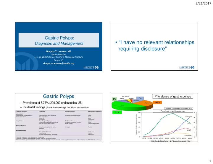
– Prevalence of 3.75% (200,000 endoscopies-US) – Incidental findings (Rare: hemorrhage / outflow obstruction)

Gastric Polyps

Prevalence of gastric polyps

FGP: Fundic Gland Polyp; GHP:Gastric Hyperplastic Polyp

77% 14.5% 7% 2%

Prevalence of gastric polyp / age

Sonnenberg A., Digestive and Liver Disease 47 (2015)

slide-2
SLIDE 2

5/26/2017 2

Sonnenberg A., Digestive and Liver Disease 47 (2015)

Fundic Gland Polyps

  • Sporadic (0.09 to 5% of endoscoped pts; Female +).
  • FAP
  • Proton pump inhibitors
  • GAPPS (Gastric Adenocarcinoma and

Proximal Polyposis)

  • Oxyntic mucosa
  • Sessile: 1-5 mm
  • Multiple (40-60%)
  • Over time 40-50% are labile
  • FAP: inactivating APC / chr 5q

allelic loss

  • Sporadic: activating β catenin

mutation (60%-90%)

slide-3
SLIDE 3

5/26/2017 3

Sporadic FGP: Dysplasia is rare (1-6%) Syndromic patients (FAP): 25-48% (LGD>HGD [0-12%]) Only 4 cases of ACA

Goddard AF. Gut 2010;59:1270-1276

  • [mean follow-up of 6 years].

– 54% stable “persisted” (n=13) – 33% “regressed” (n=8) – 13% “progressed” to HGD/IMC (n=3)

(sporadic GED progression rate: LGD: 5-14% and HGD: 24-37%)

Arnason T. Histopathology 2014,

  • Recommendations:

– Follow q. 2/3 years:

  • Look for large polyps (>1cm)
  • Sample extensively

Evolution of dysplastic FGP in FAP

slide-4
SLIDE 4

5/26/2017 4

Key features:

  • FG polyposis w/occasional

hyperplastic & adenomatous polyps,

– sparing the antrum – devt. of intestinal type GCA

  • Autosomal dominant inheritance

(Incomplete penetrance).

  • No colonic polyps
  • Point mutations in exon 1B of

APC

Gut 2012, 61:774-779 The American Journal of Human Genetics 98, 1–13, May 5, 2016

Courtesy of P. Kumarasinghe

Hyperplastic Polyps

Mean age: 65yrs -Sessile/pedunculate -Antrum:60%.

25% 12% 21% 27% 15% HP gastritis Auto Im. Gastritis

  • React. Gastro.

Others Normal

Mucosal background Multiplicity: 20%.

  • Sessile or pedunculated
  • Size is variable
slide-5
SLIDE 5

5/26/2017 5

Polypoid foveolar hyperplasia

<1cm

Thick muscle bands Thickened wall vessels Cystic glandular dilatation Glands in mid zone

Prolapse variant (of hyperplastic polyp)

208 polypoid lesions reported as hyperplastic polyps

Polypoid fov. hyperplasia:49% Hyperplastic polyp:31% Prolapse polyp:20%

slide-6
SLIDE 6

5/26/2017 6

Natural history of hyperplastic polyps

27% 68% 3% 2% Stable Disappear Growth/Disappear Increased

Dysplasia:1.8-16.4%; Carcinoma:0.3-7.1% (avg 2.1%) (> 2.0

cm)

Goddard AF. Gut 2010;59:1270-1276

Differential diagnosis of hyperplastic polyps is challenging on a superficial pinch biopsies

Hyperplastic Polyps – Diff. Dx

  • Other differential dx:

– Menetrier’s disease – Bile reflux/ post surgery gastritis – Gastritis Polyposa Cystica Juvenile polyposis Peutz-Jeghers Syndrome Cowden’s Disease Cronkhite-Canada Syndrome Inheritance autosomal dominant autosomal dominant autosomic dominant non-inherited (sporadic) Gene SMAD4 or BMPR1A STK11/LKB1 PTEN None Gastric location infrequent (15~25%) 25~50% common common Location of polyp antrum > body or fundus random random random Size of polyp variable usually small (<1cm) usually small (<1cm) variable Lifetime risk of gastric Ca. 15~20% 30% rare about 10%

slide-7
SLIDE 7

5/26/2017 7

Juvenile polyps (polyposis)

  • Median age of pts presenting with gastric polyps ~40 years
  • Rounded and sessile when small but pedunculated with a lobular

appearance as they enlarge.

Setia N. The Oncologist 2015; accepted for publication

  • No smooth muscle fibers

Disorganized pits / glands

  • f varying sizes & shapes

Disorganized pits / glands

  • f varying sizes & shapes

Pits & glands are grouped/ packeted; Unremarkable epithelium

Peutz-jeghers

Median age of Dx:16 yrs

Subtle intervening septae of smooth muscle strands Unremarkable lamina propria

slide-8
SLIDE 8

5/26/2017 8

  • Increased risk of GI cancer through the hamartoma-adenoma-

carcinoma sequence and de novo malignant change.

  • Dysplasia is noted in 2-3% of PJ polyps.

Cronkhite-Canada Syndrome

  • Protein-losing enteropathy.
  • Ectodermal changes
  • Hamartomatous polyposis.
  • (IgG4 related condition?)
  • Variable natural history

– 50-60% mortality – Electrolytes imbalance, GI bleeding, opportunistic infections

  • Malignant potential~10%
  • Broad based polyp w/ marked

stromal edema & unevenly spaced glands.

  • Mixed inflammatory

infiltrate w/ prominent eosinophilia

Gastric Adenoma

  • Nodule of gastric

dysplasia:

“Unequivocal neoplastic (non invasive) process”

  • Single in 82% of cases
  • 80-90% < 2cm
slide-9
SLIDE 9

5/26/2017 9

Architectural/Cytologic Features of Gastric Dysplasia

Glandular disarray,budding, branching and dilatation

Nuclear pleomorphism High N/C ratio Loss of nuclear polarity w/ pseudostratification Lack of differentiation w/ mucus depletion

Low Grade Dysplasia High Grade Dysplasia

Phenotypic Diversity of Gastric Dysplasia

Adenomatous Foveolar Pyloric

CD10 MUC2 MUC5AC MUC6 Intestinal (+) (Apical membrane) (+) (Goblet cells) (-) (-) Foveolar (-) (-) (+) (+/-) (glands) Pyloric (-) (-) (+) (surface) (+) (glands)

Pre-pyloric nodule

slide-10
SLIDE 10

5/26/2017 10

Polypoid gastric dysplasia, foveolar type

  • Cuboidal to low columnar cells,
  • Clear/light eosinophilic cytoplasm,
  • Round to oval nuclei.

(n=69)

Prevalence of foveolar GED: 22% (Adenomatous: 45%, hybrid 33%)

(n=69)

Muc5AC

Park DY. AJSP 2008

Grade Immunophenotype p value

Foveolar

(n=24)

Intestinal (n=22) Hybrid (n=14)

HGD (n=25) 15* (63%) 4 (18%) 6 (43%) Low grade (n=35) 9 (37%) 18 (82%) 8 (57%) 0.010 * coexistent intramucosal carcinoma in 8 cases

Foveolar differentiation is associated w/ HGD & coexistence of IMC

Valente P; Gastric Cancer 2014

  • Foveolar GED is often depressed/flat & associated w/ HGD (p= 0.046).
  • HGD associated w/ MUC5AC expression regardless of the type (p=0.026).

Type 2

MUC6 MUC5

Pyloric gland adenoma

slide-11
SLIDE 11

5/26/2017 11

MUC6

Intramucosal adenocarcinomia High Grade Dysplasia

What we know about PGA

  • Older pts (mean age: 70 years)
  • Females > males (3:1)
  • Oxyntic mucosa
  • Autoimmune gastritis +
  • FAP (no sex predominance); Lynch Sd
  • 53% with HGD (23 cases)
  • Pyloric-phenotype (MUC6+,
  • < 30% MUC5AC+)
  • GNAS mutation in 48% of cases

(none in intestinal & foveolar dysplasia)

  • KRAS mutation in 41% of cases

What is new about PGA

  • Cardia (8%), antrum (6%), pylorus

(3%)

  • 27% in AIG but 73% not, w/ 9% in

FAP & 36% in normal mucosa

  • 55% LGD [average size:1.7 cm] while

37% HGD [avg: 3.4 cm]

  • TVA architecture more common in

HGD (52%) than LGD

  • 51% coexpressed MUC5AC with

MUC6 in an intermixed pattern

  • Only 7% w/ recurrence at 1 year.

Matsubara A. J Pathol 2013;229:579

Tubular PGA with LGD

Tubulo-villous PGA with HGD

slide-12
SLIDE 12

5/26/2017 12

Middle age female

MUC6

What is your diagnosis?

  • 1. Tubular adenoma
  • 2. Well diff. adenoCA
  • 3. Fundic gland polyp

Chief cells Parietal cells

H+/K+ ATP ase

Pepsinogen I

Uyema H AJSP. 2010;609-619. Singhi A AJSP. 2012;1030-1035.

A morphologic continuum….. Anastomosing glands (55%); Mild atypia (58%) Desmoplasia (16%) Necrosis (8%)

GNAS mutation

Kushima R. Pathology International 2013

MUC6 MUC5A C

Mixed polyps have been seen

41% 48%

slide-13
SLIDE 13

5/26/2017 13

Goddard AF. Gut 2010;59:1270-1276

Gastric polyps: characteristics and management strategies NET (Neuroendocrine Tumors) in Autoimmune Gastritis

CHR SYN CD-56 Size > 0.5cm Submucosal Invasion

Gastric Neuroendocrine Tumors

Types 1,2, 3 are ECL cells tumors Type Background % Sex # of tumors Size Invasion Mets/

Prognosis

1 Autoimmune Gastritis ECL-cell hyperplasia 70 F

(65%)

Multiple

(70%) Most are small

superficial <10% Good 2 MEN-I,ZES, Hyperparathyroidism ECL-cell hyperplasia Non atrophic or hypertrophic mucosa 5-8 M=F Multiple (100%) Small

(<1cm)

superficial 10% Good 3 Sporadic Normal or chronic gastritis but no atrophy 20 M

(80%)

Single

(100%) Variable

deep 65% Mod/Poor

  • Increasing prevalence (?increase in endoscopic examination):
  • 2% of gastric malignancies (0.5% in ‘50)
  • 9% of intestinal neuroendocrine neoplasia (2.4% in ‘50)

Grade 1 (<2 mitoses x 10 HPF; <2% Ki67 index) Grade 2 (<20 mitoses x 10 HPF; 3-20% Ki67 index)

slide-14
SLIDE 14

5/26/2017 14

Type I Gastric NET

(most common)

  • Most common type (~70%)
  • Predilection for older females
  • Associated w/ autoimmune gastritis

– Hypo/achlorhydria – Hypergastrinemia & antral G-cell hyperplasia – Pernicious anemia (subset)

  • ECL cell proliferation
  • Small and multicentric
  • Rare angioinvasion
  • Metastases are exceptional
  • LN (5%); Liver mets (2.5%)

Type II Gastric NET

(least common)

  • Associated w/ ZES/MEN-1 (13-30%)
  • Hypertrophic hypersecretory

gastropathy

  • Hypergastrinemia (gastrinoma

associated)

  • ECL cell hyperplasia may be

present

  • Small and multicentric
  • Metastases are rare
  • LN (30%); Liver mets (10%)

Management type I:

  • > 2cm: high risk of metastases (resection either of the lesion or antrectomy)
  • <1cm may remain stable in most cases
  • 1-2 cm: ESD (or resection or antrectomy for numerous lesions)

Inflammatory Fibroid Polyp

  • Benign- Antrum (70%) & ileum (20%)
  • Wide age range (mean age 60)
  • Intussusception (small bowel)

Often margins Often submucosal w/ ill-defined margins Polyp (often ulcerated)> mural mass Polyp (often ulcerated)> mural mass

slide-15
SLIDE 15

5/26/2017 15

CD117

PDGFRA gain of function mutations

Schildhaus HU; J Pathol 2008;216

  • Cell of origin: fibroblastic?

Myofibroblastic?, histiocytic

  • r dendritic cells?
  • cells are (+) for CD34,

fascin and calponin, [SMA can be seen].

  • c-kit, S100, desmin:

negative.

Thank You!