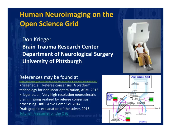
SLIDE 1 Human Neuroimaging on the Open Science Grid
Don Krieger Brain Trauma Research Center Department of Neurological Surgery University of Pittsburgh
References may be found at
https://indico.fnal.gov/contributionDisplay.py?contribId=20&sessionId=6&confId=10571
Krieger et. al., Referee consensus: A platform technology for nonlinear optimization, ACM, 2013. Krieger et. al., Very high resolution neuroelectric brain imaging realized by referee consensus
- processing. Intl J Advd Comp Sci, 2014.
Draft graphic explanation of the solver, 2015. Tutorial: Freesurfer on the OSG
SLIDE 2 Human Neuroimaging on the Open Science Grid
- 1. Concussion and functional neuroimaging, a
high value high demand use of the OSG.
SLIDE 3 David Okonkwo, Jim Becker, Sue Beers, Mickey Collins, Anthony Kontos, Malcolm McNeil, Lisa Morrow, Nora Presson, Walt Schneider
Human Neuroimaging on the Open Science Grid
- 1. Concussion and functional neuroimaging, a
high value high demand use of the OSG. Support: Dept. of Defense, Open Science Grid (NSF,DOE), XSede (NSF).
SLIDE 4 Human Neuroimaging on the Open Science Grid
- 1. Concussion and functional neuroimaging, a
high value high demand use of the OSG.
- 2. Use of Comet to boost private OSG capacity
for time sensitive job groups.
SLIDE 5 Mats Rynge, Mahidhar Tatineni, Frank Wurthwein, Miron Livny
Human Neuroimaging on the Open Science Grid
- 1. Concussion and functional neuroimaging, a
high value high demand use of the OSG.
- 2. Use of Comet to boost private OSG capacity
for time sensitive job groups. San Diego Supercomputing Center, Open Science Grid (NSF,DOE), XSede (NSF).
SLIDE 6 Human Neuroimaging on the Open Science Grid
- 1. Concussion and functional neuroimaging, a
high value high demand use of the OSG.
- 2. Use of Comet to boost private OSG capacity
for time sensitive job groups.
- 3. Freesurfer, a widely used brain image
processing package, a high value low demand use of the OSG.
SLIDE 7 Rob Gardner, Suchandra Thapa, Balamurugan Desinghu
Human Neuroimaging on the Open Science Grid
- 1. Concussion and functional neuroimaging, a
high value high demand use of the OSG.
- 2. Use of Comet to boost private OSG capacity
for time sensitive job groups.
- 3. Freesurfer, a widely used brain image
processing package, a high value low demand use of the OSG. Support: University of Chicago, Open Science Grid (NSF,DOE)
SLIDE 8
1
SLIDE 9 Human Neuroimaging on the Open Science Grid
- 1. Concussion and functional neuroimaging, a
high value high demand use of the OSG.
SLIDE 10
- Our effort is motivated by an important and
refractory clinical entity, concussion.
Concussion: TeamTBI Functional Neuroimaging
SLIDE 11
- Our effort is motivated by an important and
refractory clinical entity, concussion.
- The functional neuroimaging component of
the effort relies on MEG recording and
- analysis. We extract validated neuroelectric
events with the referee consensus solver deployed on the Open Science Grid.
Concussion: TeamTBI Functional Neuroimaging
SLIDE 12
- Our effort is motivated by an important and
refractory clinical entity, concussion.
- The functional neuroimaging component of
the effort relies on MEG recording and
- analysis. We extract validated neuroelectric
events with the referee consensus solver deployed on the Open Science Grid.
- We reduce the copious MEG‐derived
neuroelectric events to summary maps of regional brain activation.
Concussion: TeamTBI Functional Neuroimaging
SLIDE 13
- Our effort is motivated by an important and
refractory clinical entity, concussion.
- The functional neuroimaging component of
the effort relies on MEG recording and
- analysis. We extract validated neuroelectric
events with the referee consensus solver deployed on the Open Science Grid.
- We reduce the copious MEG‐derived
neuroelectric events to summary maps of regional brain activation.
- We further reduce those maps to numerical
scores which may be formally tested for relationships with clinical measures derived from symptom inventories and neuropsychological testing.
Concussion: TeamTBI Functional Neuroimaging
SLIDE 14
- Our effort is motivated by an important and
refractory clinical entity, concussion.
- The functional neuroimaging component of
the effort relies on MEG recording and
- analysis. We extract validated neuroelectric
events with the referee consensus solver deployed on the Open Science Grid.
- The data reduction steps are based on 3
simple assumptions which prove out:
Concussion: TeamTBI Functional Neuroimaging
SLIDE 15
- Our effort is motivated by an important and
refractory clinical entity, concussion.
- The functional neuroimaging component of
the effort relies on MEG recording and
- analysis. We extract validated neuroelectric
events with the referee consensus solver deployed on the Open Science Grid.
- The data reduction steps are based on 3
simple assumptions which prove out:
- 1. the brain is more widely activated during
task performance than during rest.
Concussion: TeamTBI Functional Neuroimaging
SLIDE 16
- Our effort is motivated by an important and
refractory clinical entity, concussion.
- The functional neuroimaging component of
the effort relies on MEG recording and
- analysis. We extract validated neuroelectric
events with the referee consensus solver deployed on the Open Science Grid.
- The data reduction steps are based on 3
simple assumptions which prove out:
- 1. the brain is more widely activated during
task performance than during rest.
- 2. The rate at which validated neuroelectric
events occur within a given volume increases with activation.
Concussion: TeamTBI Functional Neuroimaging
SLIDE 17
- Our effort is motivated by an important and
refractory clinical entity, concussion.
- The functional neuroimaging component of
the effort relies on MEG recording and
- analysis. We extract validated neuroelectric
events with the referee consensus solver deployed on the Open Science Grid.
- The data reduction steps are based on 3
simple assumptions which prove out:
- 1. the brain is more widely activated during
task performance than during rest.
- 2. The rate at which validated neuroelectric
events occur within a given volume increases with activation.
- 3. The corresponding averaged current
amplitude also increases with activation.
Concussion: TeamTBI Functional Neuroimaging
SLIDE 18
Inside the liquid helium dewar is an array of 102 1”x1” chips, each with 3 magnetic field sensors. Hence the 3D “shape” of the magnetic field around the head is sampled at 102 points .
SLIDE 19
This 3D snapshot of the “shape” of the magnetic field is obtained 1000 times per second. Each snapshot is a set of 306 measurements at a particular time and is designated: Mt
SLIDE 20
This 3D snapshot of the “shape” of the magnetic field is obtained 1000 times per second. Each snapshot is a set of 306 measurements at a particular time and is designated: Mt The referee consensus solver uses 80 snapshots at a time.
SLIDE 21
Given a specific location in the brain, X, the solver determines if an electric current at X is contributing significantly (p < 10‐12) to the shape of the magnetic field.
SLIDE 22
Given a specific location in the brain, X, the solver determines if an electric current at X is contributing significantly (p < 10‐12) to the shape of the magnetic field. As part of this determination the procedure produces a high fidelity estimate of the time course of the electric current at X.
SLIDE 23 For each validated current source event, we have
- time marker … 1 msec resolution
- 3D location … ≈ 1 mm resolution
- 3D direction … 2 components: The radial component cannot be
estimated.
- 80 msec waveform, i.e. the time course of the amplitude of the current
SLIDE 24 For each validated current source event, we have
- time marker … 1 msec resolution
- 3D location … ≈ 1 mm resolution
- 3D direction … 2 components: The radial component cannot be
estimated.
- 80 msec waveform, i.e. the time course of the amplitude of the current
1‐15 sources per cm3 per second are typically found.
SLIDE 25 For each validated current source event, we have
- time marker … 1 msec resolution
- 3D location … ≈ 1 mm resolution
- 3D direction … 2 components: The radial component cannot be
estimated.
- 80 msec waveform, i.e. the time course of the amplitude of the current
1‐15 sources per cm3 per second are typically found. For a typical 1.5 liter brain this represents 1000‐15,000 events/sec , i.e. 1,800,000‐27,000,000 events from a ½ hour recording session.
SLIDE 26 For each validated current source event, we have
- time marker … 1 msec resolution
- 3D location … ≈ 1 mm resolution
- 3D direction … 2 components: The radial component cannot be
estimated.
- 80 msec waveform, i.e. the time course of the amplitude of the current
1‐15 sources per cm3 per second are typically found. For a typical 1.5 liter brain this represents 1000‐15,000 events/sec , i.e. 1,800,000‐27,000,000 events from a ½ hour recording session. The stringent acceptance criterion, p < 10‐12 for each accepted source, insures high confidence that all identified sources are real.
SLIDE 27 For each validated current source event, we have
- time marker … 1 msec resolution
- 3D location … ≈ 1 mm resolution
- 3D direction … 2 components: The radial component cannot be
estimated.
- 80 msec waveform, i.e. the time course of the amplitude of the current
1‐15 sources per cm3 per second are typically found. For a typical 1.5 liter brain this represents 1000‐15,000 events/sec , i.e. 1,800,000‐27,000,000 events from a ½ hour recording session. The stringent acceptance criterion, p < 10‐12 for each accepted source, insures high confidence that all identified sources are real. Referee consensus processed MEG recordings yield an accepted source approximately 1/1500th of the time.
SLIDE 28 For each validated current source event, we have
- time marker … 1 msec resolution
- 3D location … ≈ 1 mm resolution
- 3D direction … 2 components: The radial component cannot be
estimated.
- 80 msec waveform, i.e. the time course of the amplitude of the current
1‐15 sources per cm3 per second are typically found. For a typical 1.5 liter brain this represents 1000‐15,000 events/sec , i.e. 1,800,000‐27,000,000 events from a ½ hour recording session. The stringent acceptance criterion, p < 10‐12 for each accepted source, insures high confidence that all identified sources are real. Referee consensus processed MEG recordings yield an accepted source approximately 1/1500th of the time. In principle comparable measurement resolution could be obtained with an array of 1,500,000 indwelling low impedance wires implanted in the brain on a 1 mm grid. That direct but unrealizable approach would extract more information since it would yield continuous recordings from each location.
SLIDE 29
Each identified electrical current is flowing in a direction. These typical neuroelectric current events were found in a 1 second MEG recording in a 1 mm thick volume.
rest task 373 current sources 387 current sources
SLIDE 30
For each volunteer, we compare the activation of each cortical gray matter region with that of the adjacent white matter rim.
Differential Activation – Gray/White
Gray > White (green) White > Gray (red)
SLIDE 31 For each volunteer, we compare the activation of each cortical gray matter region with that of the adjacent white matter rim.
- 34 gray/white pairs within each brain hemisphere
- p‐value < 10‐6 for each accepted gray/white pair
Differential Activation – Gray/White
Gray > White (green) White > Gray (red)
SLIDE 32 For each volunteer, we compare the activation of each cortical gray matter region with that of the adjacent white matter rim.
- 34 gray/white pairs within each brain hemisphere
- p‐value < 10‐6 for each accepted gray/white pair
For most pairs the white matter is more strongly activated than the gray. This result is both unprecedented …
Differential Activation – Gray/White
Gray > White (green) White > Gray (red)
SLIDE 33 For each volunteer, we compare the activation of each cortical gray matter region with that of the adjacent white matter rim.
- 34 gray/white pairs within each brain hemisphere
- p‐value < 10‐6 for each accepted gray/white pair
For most pairs the white matter is more strongly activated than the gray. This result is both unprecedented and robust.
Differential Activation – Gray/White
Gray > White (green) White > Gray (red) Norm
SLIDE 34 For each volunteer, we compare the activation of each cortical gray matter region with that of the adjacent white matter rim.
- 34 gray/white pairs within each brain hemisphere
- p‐value < 10‐6 for each accepted gray/white pair
For most pairs the white matter is more strongly activated than the gray. This result is both unprecedented and robust. No comparable result is currently obtainable from any neuroimaging modality processed in any other way.
Differential Activation – Gray/White
Gray > White (green) White > Gray (red) Norm
SLIDE 35
2
SLIDE 36 Human Neuroimaging on the Open Science Grid
- 1. Concussion and functional neuroimaging, a
high value high demand use of the OSG.
- 2. Use of Comet to boost private OSG capacity
for time sensitive job groups.
SLIDE 37
Monday – Wednesday: Each TeamTBI volunteer is evaluated with a battery of neuroimage, neuropsychological, vestibular, ocular, and sleep tests.
Concussion: TeamTBI Functional Neuroimaging
SLIDE 38
Monday – Wednesday: Each TeamTBI volunteer is evaluated with a battery of neuroimage, neuropsychological, vestibular, ocular, and sleep tests. Thursday morning: All results are presented and discussed in an adjudication conference.
Concussion: TeamTBI Functional Neuroimaging
SLIDE 39
Monday – Wednesday: Each TeamTBI volunteer is evaluated with a battery of neuroimage, neuropsychological, vestibular, ocular, and sleep tests. Thursday morning: All results are presented and discussed in an adjudication conference. This provides a narrow time window (36‐60 hr) to process the MEG recordings, ≈ 90,000 OSG hr.
Concussion: TeamTBI Functional Neuroimaging
SLIDE 40
Monday – Wednesday: Each TeamTBI volunteer is evaluated with a battery of neuroimage, neuropsychological, vestibular, ocular, and sleep tests. Thursday morning: All results are presented and discussed in an adjudication conference. This provides a narrow time window (36‐60 hr) to process the MEG recordings, ≈ 90,000 OSG hr.
Concussion: TeamTBI Functional Neuroimaging
To support these time sensitive runs, XSede has allocated 50,000 SUs on Comet per MEG study.
SLIDE 41
Monday – Wednesday: Each TeamTBI volunteer is evaluated with a battery of neuroimage, neuropsychological, vestibular, ocular, and sleep tests. Thursday morning: All results are presented and discussed in an adjudication conference. This provides a narrow time window (36‐60 hr) to process the MEG recordings, ≈ 90,000 OSG hr.
Concussion: TeamTBI Functional Neuroimaging
To support these time sensitive runs, XSede has allocated 50,000 SUs on Comet per MEG study. No guarantee is needed or provided.
SLIDE 42
Monday – Wednesday: Each TeamTBI volunteer is evaluated with a battery of neuroimage, neuropsychological, vestibular, ocular, and sleep tests. Thursday morning: All results are presented and discussed in an adjudication conference. This provides a narrow time window (36‐60 hr) to process the MEG recordings, ≈ 90,000 OSG hr.
Concussion: TeamTBI Functional Neuroimaging
To support these time sensitive runs, XSede has allocated 50,000 SUs on Comet per MEG study. No guarantee is needed or provided. When added to the SUs we get from the opportunistic pool, this enables sufficient processing to provide reliable results for adjudication.
SLIDE 43
Restrict opportunistic jobs to run on non‐Comet IP addresses.
Comet booster for OSG condor pool
.
SLIDE 44 Restrict opportunistic jobs to run on non‐Comet IP addresses. Comet jobs:
- 1. Restrict OSG jobs to Comet IP addresses.
Assign Non‐XSede account number.
Comet booster for OSG condor pool
.
SLIDE 45 Restrict opportunistic jobs to run on non‐Comet IP addresses. Comet jobs:
- 1. Restrict OSG jobs to Comet IP addresses.
Assign Non‐XSede account number.
Comet booster for OSG condor pool
.
SLIDE 46 Restrict opportunistic jobs to run on non‐Comet IP addresses. Comet jobs:
- 1. Restrict OSG jobs to Comet IP addresses.
Assign Non‐XSede account number.
- 2. Queue the OSG jobs.
- 3. Spawn glideins on Comet. The glideins will
fall under XSede accounting.
Comet booster for OSG condor pool
.
SLIDE 47
Comet booster for OSG condor pool Live Website
Blue line … # Jobs Running
SLIDE 48
Comet booster for OSG condor pool Live Website
Blue line … # Jobs Running Blue dots … # Comet Glidein Job Slots
SLIDE 49
Comet booster for OSG condor pool Live Website
Blue line … # Jobs Running Blue dots … # Comet Glidein Job Slots 2 3‐hour development runs.
SLIDE 50
Comet booster for OSG condor pool Live Website
Blue line … # Jobs Running Blue dots … # Comet Glidein Job Slots 2 3‐hour development runs. The live monitor was developed after starting the first run.
SLIDE 51
Comet booster for OSG condor pool Live Website
Blue line … # Jobs Running Blue dots … # Comet Glidein Job Slots 2 3‐hour development runs. The live monitor was developed after starting the first run. MIN_UNTIL_RETIREMENT was adjusted for the second run.
SLIDE 52
Comet booster for OSG condor pool Live Website
Blue line … # Jobs Running Blue dots … # Comet Glidein Job Slots
SLIDE 53
Comet booster for OSG condor pool Live Website
Blue line … # Jobs Running Blue dots … # Comet Glidein Job Slots 30‐hour production run – 2 MEG’s
SLIDE 54
Comet booster for OSG condor pool Live Website
Blue line … # Jobs Running Blue dots … # Comet Glidein Job Slots 30‐hour production run – 2 MEG’s Opportunistic hours were high for 12 hours.
SLIDE 55
3
SLIDE 56 Human Neuroimaging on the Open Science Grid
- 1. Concussion and functional neuroimaging, a
high value high demand use of the OSG.
- 2. Use of Comet to boost private OSG capacity
for time sensitive job groups.
- 3. Freesurfer, a widely used brain image
processing package, a high value low demand use of the OSG.
SLIDE 57 Freesurfer on the Open Science Grid
Access to the OSG is open to universities, government labs, many
57
SLIDE 58 Freesurfer on the Open Science Grid
Access to the OSG is open to universities, government labs, many
Your need must be sufficient to motivate your start‐up effort. The great majority of potential users have a modest need with consequently limited motivation.
58
SLIDE 59 Freesurfer on the Open Science Grid
Access to the OSG is open to universities, government labs, many
Your need must be sufficient to motivate your start‐up effort. The great majority of potential users have a modest need with consequently limited motivation. To reduce the start‐up effort we are trying a simple approach:
- Provide turn‐key command line tools for one software package
at a time.
59
SLIDE 60 Freesurfer on the Open Science Grid
Access to the OSG is open to universities, government labs, many
Your need must be sufficient to motivate your start‐up effort. The great majority of potential users have a modest need with consequently limited motivation. To reduce the start‐up effort we are trying a simple approach:
- Provide turn‐key command line tools for one software package
at a time.
- We have selected a brain image processing application as a
demonstration, Freesurfer (Martinos Biomedical Imaging Center, Harvard University).
60
SLIDE 61 Freesurfer on the Open Science Grid
The primary function of Freesurfer is readily adapted for execution on the Open Science Grid:
- Each job is fully independent.
61 Cortical gray matter (green) vs adjacent white matter (red) differential neuroelectric activation. Regions identified with freesurfer.
SLIDE 62 Freesurfer on the Open Science Grid
The primary function of Freesurfer is readily adapted for execution on the Open Science Grid:
- Each job is fully independent.
- Execution time: 16‐24 hours (single core), 4‐8 hours (multiple cores)
62 Cortical gray matter (green) vs adjacent white matter (red) differential neuroelectric activation. Regions identified with freesurfer.
SLIDE 63 Freesurfer on the Open Science Grid
The primary function of Freesurfer is readily adapted for execution on the Open Science Grid:
- Each job is fully independent.
- Execution time: 16‐24 hours (single core), 4‐8 hours (multiple cores)
- Memory requirement: ~3 GBytes.
63 Cortical gray matter (green) vs adjacent white matter (red) differential neuroelectric activation. Regions identified with freesurfer.
SLIDE 64 Freesurfer on the Open Science Grid
The primary function of Freesurfer is readily adapted for execution on the Open Science Grid:
- Each job is fully independent.
- Execution time: 16‐24 hours (single core), 4‐8 hours (multiple cores)
- Memory requirement: ~3 GBytes.
- Input to each job: ~10 MBytes.
64 Cortical gray matter (green) vs adjacent white matter (red) differential neuroelectric activation. Regions identified with freesurfer.
SLIDE 65 Freesurfer on the Open Science Grid
The primary function of Freesurfer is readily adapted for execution on the Open Science Grid:
- Each job is fully independent.
- Execution time: 16‐24 hours (single core), 4‐8 hours (multiple cores)
- Memory requirement: ~3 GBytes.
- Input to each job: ~10 MBytes.
- Output from each job: ~400 MBytes.
65 Cortical gray matter (green) vs adjacent white matter (red) differential neuroelectric activation. Regions identified with freesurfer.
SLIDE 66 Freesurfer on the Open Science Grid
The primary function of Freesurfer is readily adapted for execution on the Open Science Grid:
- Each job is fully independent.
- Execution time: 16‐24 hours (single core), 4‐8 hours (multiple cores)
- Memory requirement: ~3 GBytes.
- Input to each job: ~10 MBytes.
- Output from each job: ~400 MBytes.
Freesurfer is a high‐value low‐demand use of the Open Science Grid.
66 Cortical gray matter (green) vs adjacent white matter (red) differential neuroelectric activation. Regions identified with freesurfer.
SLIDE 67 Freesurfer on the Open Science Grid
The primary function of Freesurfer is readily adapted for execution on the Open Science Grid:
- Each job is fully independent.
- Execution time: 16‐24 hours (single core), 4‐8 hours (multiple cores)
- Memory requirement: ~3 GBytes.
- Input to each job: ~10 MBytes.
- Output from each job: ~400 MBytes.
Freesurfer is a high‐value low‐demand use of the Open Science Grid. Many Freesurfer users lack sufficient computing resources and/or software expertise to install and maintain the package.
67 Cortical gray matter (green) vs adjacent white matter (red) differential neuroelectric activation. Regions identified with freesurfer.
SLIDE 68 Freesurfer on the Open Science Grid
The primary function of Freesurfer is readily adapted for execution on the Open Science Grid:
- Each job is fully independent.
- Execution time: 16‐24 hours (single core), 4‐8 hours (multiple cores)
- Memory requirement: ~3 GBytes.
- Input to each job: ~10 MBytes.
- Output from each job: ~400 MBytes.
Freesurfer is a high‐value low‐demand use of the Open Science Grid. Many Freesurfer users lack sufficient computing resources and/or software expertise to install and maintain the package. There is a user base of at least 3,000 world‐wide.
68 Cortical gray matter (green) vs adjacent white matter (red) differential neuroelectric activation. Regions identified with freesurfer.
SLIDE 69 Freesurfer on the Open Science Grid
Our demonstration effort has produced turn‐key command line tools usable either from the osgconnect remote client or from the osgconnect submit host:
69
SLIDE 70 Freesurfer on the Open Science Grid
Our demonstration effort has produced turn‐key command line tools usable either from the osgconnect remote client or from the osgconnect submit host:
- Submit a Freesurfer job to the grid.
70 fsurf –submit –subject MRN_1 –dir INPUT_DIRECTORY ;# 4-8 hours fsurf –submit –subject MRN_1 –dir INPUT_DIRECTORY –dualcore ;# 11-16 hours
SLIDE 71 Freesurfer on the Open Science Grid
Our demonstration effort has produced turn‐key command line tools usable either from the osgconnect remote client or from the osgconnect submit host:
- Submit a Freesurfer job to the grid.
- Monitor job status.
71 fsurf –submit –subject MRN_1 –dir INPUT_DIRECTORY ;# 4-8 hours fsurf –submit –subject MRN_1 –dir INPUT_DIRECTORY –dualcore ;# 11-16 hours fsurf –list fsurf –status –subject MRN_1
SLIDE 72 Freesurfer on the Open Science Grid
Our demonstration effort has produced turn‐key command line tools usable either from the osgconnect remote client or from the osgconnect submit host:
- Submit a Freesurfer job to the grid.
- Monitor job status.
- Fetch the results when complete.
72 fsurf –submit –subject MRN_1 –dir INPUT_DIRECTORY ;# 4-8 hours fsurf –submit –subject MRN_1 –dir INPUT_DIRECTORY –dualcore ;# 11-16 hours fsurf –list fsurf –status –subject MRN_1 fsurf –output –subject MRN_1 fsurf –remove –subject MRN_1
SLIDE 73 Freesurfer on the Open Science Grid
Benchmark testing has been completed for several data flows and for a range of core counts.
73 fsurf –submit –subject MRN_1 –dir INPUT_DIRECTORY ;# 4-8 hours fsurf –submit –subject MRN_1 –dir INPUT_DIRECTORY –dualcore ;# 11-16 hours fsurf –list fsurf –status –subject MRN_1 fsurf –output –subject MRN_1 fsurf –remove –subject MRN_1
SLIDE 74 Freesurfer on the Open Science Grid
Benchmark testing has been completed for several data flows and for a range of core counts. Results will be validated for the range of RHEL versions running on the OSG including a very early and very recent one.
74 fsurf –submit –subject MRN_1 –dir INPUT_DIRECTORY ;# 4-8 hours fsurf –submit –subject MRN_1 –dir INPUT_DIRECTORY –dualcore ;# 11-16 hours fsurf –list fsurf –status –subject MRN_1 fsurf –output –subject MRN_1 fsurf –remove –subject MRN_1
SLIDE 75 Freesurfer on the Open Science Grid
Benchmark testing has been completed for several data flows and for a range of core counts. Results have been validated for the range of RHEL versions running on the OSG including a very early and very recent one. Alpha user testing is complete. Beta user testing and feedback will be by computer support people with little or no knowledge of the project.
75 fsurf –submit –subject MRN_1 –dir INPUT_DIRECTORY ;# 4-8 hours fsurf –submit –subject MRN_1 –dir INPUT_DIRECTORY –dualcore ;# 11-16 hours fsurf –list fsurf –status –subject MRN_1 fsurf –output –subject MRN_1 fsurf –remove –subject MRN_1
SLIDE 76 Freesurfer on the Open Science Grid
Benchmark testing has been completed for several data flows and for a range of core counts. Results have been validated for the range of RHEL versions running on the OSG including a very early and very recent one. Alpha user testing is complete. Beta user testing and feedback will be by computer support people with little or no knowledge of the project. Final testing and feedback will be by members of the Freesurfer group.
76 fsurf –submit –subject MRN_1 –dir INPUT_DIRECTORY ;# 4-8 hours fsurf –submit –subject MRN_1 –dir INPUT_DIRECTORY –dualcore ;# 11-16 hours fsurf –list fsurf –status –subject MRN_1 fsurf –output –subject MRN_1 fsurf –remove –subject MRN_1
SLIDE 77 Human Neuroimaging on the Open Science Grid
- 1. Concussion and functional neuroimaging, a
high value high demand use of the OSG.
- 2. Use of Comet to boost private OSG capacity
for time sensitive job groups.
- 3. Freesurfer, a widely used brain image
processing package, a high value low demand use of the OSG.