help what do i do with those granulomas in the lung
play

Help! What do I do with those granulomas in the lung? 2019 Anatomic - PDF document

Help! What do I do with those granulomas in the lung? 2019 Anatomic Pathology Update University of Utah Park City, Utah Henry D. Tazelaar, M.D. Chair and Geraldine Zeiler Colby Professor of Cytopathology Department of Laboratory Medicine and


  1. Help! What do I do with those granulomas in the lung? 2019 Anatomic Pathology Update University of Utah Park City, Utah Henry D. Tazelaar, M.D. Chair and Geraldine Zeiler Colby Professor of Cytopathology Department of Laboratory Medicine and Pathology Alix College of Medicine and Science Mayo Clinic Arizona Objectives/Outline At the end of the lecture, participants should be able to… • Provide a framework for approaching cases with granulomatous inflammation  Large granulomas  Small granulomas • List the features of granulomas associated with infection • List the features of granulomas which favor a vasculitic process • Discriminate between foreign material and 
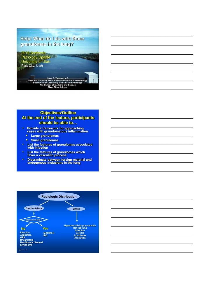
endogenous inclusions in the lung Radiologic Distribution Focal/Multi-Focal Diffuse Bronchiectasis ? Hypersensitivity pneumonitis Yes Hot tub lung No Infection Infection MAC/MLS Sarcoid Aspiration IBD Lymphoma GPA Aspiration Rheumatoid Nec/Nodular Sarcoid Lymphoma

  2. Granulomas in Biopsy Necrotizing Non-necrotizing Small Gross nodule(s) Small Gross nodule(s) Nodular Infection Infection HP Aspiration Hot tub sarcoid Sarcoid GPA Infection Lymphoma Drug Rheumatoid Lymphoma Nec Sarcoid LIP History • A 55 yr old man was found to have 2 lung nodules • History: intercapillary glomerulosclerosis • Underwent surgical lung biopsy

  3. Diagnosis?

  4. Diagnosis? Necrotizing Granulomatous Inflammation most c/w an Infectious Etiology Additional Studies • Serologies including pANCA and cANCA were negative • Stains for acid fast neg • Stains for fungi… Diagnosis Histoplasmosis characterized by Necrotizing Granulomatous Inflammation and Vasculitis

  5. Key Histologic Features • Granulomas – Necrotizing – Round borders – Geographic borders – Non-necrotizing – Surrounded by thin rim of infl’n – Some bronchiolocentric • Vasculitis • Presence of calcified bodies Key Histologic Features • Granulomas – Necrotizing – Round borders – Geographic borders – Non-necrotizing – Surrounded by thin rim of infl’n – Some bronchiolocentric • Vasculitis • Presence of calcific inclusions Solitary Granulomas Culture Results for Histoplasma Cases Source N, Pos/total Sputum 0/22 Bronchial Wash 0/9 Bronchial Brush 0/3 Needle Aspiration 0/2 Lung Biopsy 0/19 Ulbright and Katzenstein, Am J Surg Pathol 1980; 4:13-28

  6. % Yield of Cultures (C) vs. Histology (H) 80 88 Cases 70 10 Centers 60 (7 Countries) 50 Fungi 40 Mycobacteria 30 20 Mycobacteria more often culture positive 10 Fungi, more often 0 histology positive C+ H- H+ C- Both + Mukhopadhyay S et al. J Clin Pathol 2012; 65:51-7 Solitary Granulomas Importance of Special Stains • Among all patients with histoplasmosis, 54 blocks were stained with GMS • Only 74% contained organisms • Organisms “numerous” 58% – Usually located centrally Ulbright and Katzenstein, Am J Surg Pathol 1980; 4:13-28 Histol’ic features of Histoplasmosis forming a Solitary Nodule, N=24 Feature % Round borders 79 Geog’ic borders 21 Ulbright and Katzenstein, Am J Surg Pathol 1980; 4:13-28

  7. Histol’ic features of Histoplasmosis forming a Solitary Nodule, N=24 Feature % Non-nec gran 13 Vasculitis 54 Ulbright and Katzenstein, Am J Surg Pathol 1980; 4:13-28

  8. Histo – Well Formed Non-Nec Gran Acute Histoplasmosis Mukhopadhyay S et at Am J Surg Pathol 2010; 43:541-546 Endogenously-Derived Crystals Ca oxalate Ca carbonate Visscher D et al Mod Pathol 1988;1:415

  9. Aspirated Material Ca carbonate Differential Diagnosis • Other necrotizing granulomatous infections • Wegener granulomatosis /Granulomatosis with polyangiitis • Catheter sheath emboli Mycobacterium tuberculosis Histologic Feature % – Vasculitis 87 – Geographic necrosis 30 – Non-caseating granulomas 30 Ulbright and Katzenstein, Am J Surg Pathol 1980; 4:13-28

  10. TB with vasculitis Pneumocystis jivorecii Granulomatous PCP 20 Cases Disease % HIV 35 Heme malig. 30 Solid tumor 20 Unknown 15 Hartel PH et al Am J Surg Pathol 2010; 34:730-4

  11. Granulomatous PCP 20 Cases Feature % Necrotizing gran 80 Non necrotizing gran 20 Foamy exudate 25 Cystic spaces 5 Calcification 5 Hartel PH et al Am J Surg Pathol 2010; 34:730-4 GMS

  12. HISTO PCP Infectious Granulomas vs Vasculitis • Infection favored – Non-necrotizing granulomas – Sarcoid-like granulomas – Thin rim of inflammation – Giant cells that contain inclusions • Diagnosis of infection may take – Repeating special stains – Doing stains on more blocks Vasculitis and Geographic Necrosis not helpful Infectious Granulomas vs Vasculitis • Vasculitis favored - Only necrotizing granulomas - All granulomas have geographic necrosis - Granulomas/necrosis set in inflammatory background - Microabscess-like foci - Bizarre hyperchromatic nuclei in GC’s - NO inclusions in GC’s - Foci of alveolar hemorrhage or capillaritis

  13. Wegener Granulomatosis /Granulmatosis with Polyangiitis • May present as solitary pul nodule • cANCA negative in ~ 30% of patients with limited disease Pulmonary Sarcoidosis Histology • Granulomatous inflammation – Lymphangitic – Well-formed – Often hyalinized • Rare – Isolated giant cells – Interstitial and organizing pneumonia

  14. Sarcoidosis-lymphangitic Sarcoidosis-lymphangitic Lack of interstitial pneumonia

  15. Granulomas well- formed and with hyalined fibrosis Granulomas well- formed and with hyalinized fibrosis Sarcoid : Features • Non-necrotizing granulomatous inflammation without – Chronic interstitial pneumonia – Organizing pneumonia • Inclusions usually endogenous

  16. Pulmonary Sarcoid Differential Diagnosis • Hypersensitivity pneumonitis – Hot tub lung • Infection – MAC: bronchiectasis • Berylliosis Hypersensitivity Pneumonitis Diagnosis? • Exposure history? only in 50% • Antibodies testing? – Antibodies not available for many antigens – Many “exposed” but not ill patients antibodies + – Currently NOT recommended in work up Hypersensitivity Pneumonitis Diagnosis? • A difficult clinical challenge • Compatible clinical, radiographic or physiologic findings • BAL with lymphocytosis (low CD4:8) • Histopathology

  17. Hypersensitivity Pneumonitis Clinical Presentations* • Acute: dyspnea, cough, myalgias, chills etc. – 2- 9 hours after exposure – Resolves without specific therapy • Subacute/Chronic: dyspnea, cough, weight loss, anorexia *depends on intensity and duration of exposure Extrinsic Allergic Alveolitis Hypersensitivity Pneumonitis Histologic Features • Non-nec granulomatous inflamm and giant cells – Airway-centered and random – Interstitial and airspace • Interstitial pneumonia • Chronic bronchiolitis Castonguay M et al Human Pathol 2015;46:607-13l

  18. Hypersensitivity Pneumonitis Variable Histologic Features • Prominent centrilobular airspace foam cells • Organizing pneumonia • Interstitial fibrosis • Honeycomb change/ UIP-like features Eosinophils uncommon Only 20%

  19. Major Histologic Patterns in HP n=110 (%) Cell. F NSIP UIP Peribronchiolar Bronchiolocentric NSIP inflamm with fibrosis grans 45 20 6 20 9 • 19% of cases had no granulomas or giant cells • Granulomas and giant often absent in cases with fibrosis Wang p et al. Chest 2017;152:502-509

  20. Patient had 300 BIRDS in his house! Chronic hypersensitivity pneumonitis in patients diagnosed with idiopathic pulmonary fibrosis • 20/46 pts with IPF according to 2011 ATS/ERS guidelines were subsequently diagnosed with chronic HP • Bronchial challenge, lung biopsy and serum precipitins • Most attributed to occult avian antigen exposure from… down bedding Morell F et al. Lancet Resp Med 2013;1:685-94

  21. Clues to the Diagnosis of HP Feature Pattern UIP NSIP Mosaicism/air ++ ++ trapping Granulomas and ++ ++ giant cells Bronchiolocentric ++ ++ inflammation Peribronchiolar ++ + metaplasia Bridging fibrosis + + Bridging fibrosis Churg A et al Arch Pathol Lab Med 2018;142:109-119 Chronic HP with UIP pattern Prominent peribronchiolar metaplasia

  22. CHP with UIP vs IPF Does the diagnosis matter? • Differences in survival-IPF still worse in most studies • Differences in treatment-possible benefit of antigen avoidance • Differences post lung transplant- CHP may do better than IPF patients Chronic Hypersensitivity Pneumonitis • Increasingly recognized as a cause for non-fibrotic and fibrotic diffuse lung disease • Still no gold standard for diagnosis • Must always be in the differential diagnosis for UIP and NSIP patterns • Subtle clues “Hot Tub” Lung • Granulomatous lung disease due to exposure to water contaminated with atypical mycobacteria (MAC) • Flu-like illness after exposure • CT: Interstitial and nodular miliary infiltrates • Other water sources: Showers, faucets, saunas

  23. Middle Lobe Fibrocavitary Syndrome Disease Bronchiectasis MAC-Related Lung Disease IC Host Hot Tub HIV/AIDS Lung

  24. MAC: Evaluation with CT • N=62 • 56 % intact immune system • Nodular infiltrates • Bronchiectasis Hartman TE et al. Radiology, 1993; 187:3-6 Atypical Mycobacterial Infections with Bronchiectasis/ Airway Disease • Nec and non-nec. granulomas + Airway disease +/- Interstitial pneumonia MAC-Airway Disease

  25. MAC-Airway Disease MAC-Airway Disease MAC-Airway Disease

Download Presentation
Download Policy: The content available on the website is offered to you 'AS IS' for your personal information and use only. It cannot be commercialized, licensed, or distributed on other websites without prior consent from the author. To download a presentation, simply click this link. If you encounter any difficulties during the download process, it's possible that the publisher has removed the file from their server.

Recommend


More recommend