Fresh Osteochondral Allografts: The Gold Standard for Focal Femoral - - PowerPoint PPT Presentation

fresh osteochondral allografts the gold standard for
SMART_READER_LITE
LIVE PREVIEW

Fresh Osteochondral Allografts: The Gold Standard for Focal Femoral - - PowerPoint PPT Presentation

Fresh Osteochondral Allografts: The Gold Standard for Focal Femoral Condyle Lesions William Bugbee, MD Scripps Clinic Shiley Center for Orthopaedic Research and Education La Jolla, CA Disclosure Joint Restoration Foundation


slide-1
SLIDE 1

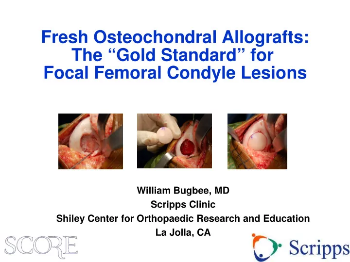
Fresh Osteochondral Allografts: The “Gold Standard” for Focal Femoral Condyle Lesions

William Bugbee, MD Scripps Clinic Shiley Center for Orthopaedic Research and Education La Jolla, CA

slide-2
SLIDE 2

Disclosure

  • Joint Restoration Foundation

– Consultant, research support

  • Arthrex

– Consultant

  • I have no IP related to osteochondral allografts

– Everything I know is in the public domain

  • I have nothing against cell based therapy or any
  • ther cartilage restoration technique
slide-3
SLIDE 3

Two Fundamental Strategies of Cartilage Restoration

  • Cell based

– Induce cells to form (chondral) tissue in situ » Marrow stimulation/ MFx plus » (M)ACI » Minced tissue (alive or dead)

  • Whole tissue based

– Restore defect with mature tissue » Osteochondral autograft (OAT) » Osteochondral allograft (OCA) » Processed (acellular) allografts

slide-4
SLIDE 4

“Seed vs. Sod”

slide-5
SLIDE 5

Subchondral Bone and the Osteochondral Unit

  • Integrated organ system
  • Interdependent structure-

function relationship

  • Increasingly recognized in

cartilage injury and repair

slide-6
SLIDE 6

28 year old Basketball Player

2cm2 MFC lesion, Previous meniscectomy

slide-7
SLIDE 7

Osteochondral Allografting

  • Originally introduced as a joint reconstructive

procedure for trauma, tumors and arthritis

  • Now widely used as a cartilage restoration technique

for chondral and osteochondral lesions

Cartilage repair paradigm

  • Microfracture
  • OAT
  • ACI
  • OCA

Complex reconstruction paradigm

  • OCD
  • AVN
  • Post-traumatic
slide-8
SLIDE 8

Clinical Outcomes Depend on Diagnosis

Diagnosis OCA failure Mean IKDC pain Mean IKDC Function Satisfaction* Traumatic chondral injury 2% 3.3 7.3 90% Osteochondritis dissecans 7% 2.1 8.1 96% Fracture 15% 4.4 6.1 80% Degenerative chondral lesion 21% 3.7 6.3 81% Avascular necrosis 25% 2.7 7.1 92% Osteoarthritis 39% 3.5 5.8 79% Among patients with grafts in situ at latest follow-up

*responded either “satisfied” or “extremely satisfied”

slide-9
SLIDE 9

Osteochondral Allograft (OCA) “Modern Technique”

  • “Typical” cartilage repair indications
  • Traumatic chondral lesions,

degenerative chondral lesions, OCD

  • Single defect
  • Precision surgical instruments
  • Dowel allografts utilizing the minimum

amount of bone needed for fixation

Technically easier to perform than shell grafts Bone transplantation kept to a minimum Fixation generally not required

Advantages

slide-10
SLIDE 10

Surgical Technique: Femoral Condyle

slide-11
SLIDE 11

Surgical Technique: Femoral Condyle

slide-12
SLIDE 12

Surgical Technique: Femoral Condyle

slide-13
SLIDE 13

1983 – present N = 1,008 Single surgeon 1997 - present N = 557 ≥ 2 years from surgery N = 225 Primary knee OCA N = 744 Minimum 2 year follow-up N = 200

Patient Population

Met inclusion criteria N = 275 Exclusions

  • Diagnosis of avascular necrosis
  • Anatomical location other than

femoral condyle

  • Grafts located on medial and lateral

femoral condyle in same knee

  • More than 2 grafts used
  • Shell grafts

200 knees (187 patients) 1999 - 2014

slide-14
SLIDE 14

Demographics

  • Average age 31 years (range, 11 – 67)
  • 63% male
  • 86% had previous surgery on operative knee
  • Median 2 previous surgeries (range, 1 – 13)

63% 23% 14%

Osteochondritis dissecans Degenerative Traumatic

  • Diagnosis
slide-15
SLIDE 15

Graft Details

  • Femoral condyle location

Medial (69%) Lateral (31%)

  • Mean total graft area 6.3 cm2

(range, 2.3 – 13)

  • Mean graft thickness 6.5 mm

(range, 5 – 11)

  • Number of grafts

1 (73%) 2 (27%)

slide-16
SLIDE 16

Subjective Outcomes

slide-17
SLIDE 17

Results: Reoperations

Reoperations 26% (52 of 200 knees) Reoperations 26% (52 of 200 knees) Not related to allograft 18% (36 of 200 knees)

Examples: Diagnostic arthroscopy Debridement Loose body removal Plate/screw removal Meniscus repair Osteotomy

Reoperations 26% (52 of 200 knees) Allograft failure 8% (16 of 200 knees)

Allograft revision (4 knees) Arthrosurface (1 knee) Uni knee arthroplasty (6 knees) Total knee arthroplasty (5 knees)

Not related to allograft 18% (36 of 200 knees)

Examples: Diagnostic arthroscopy Debridement Loose body removal Plate/screw removal Meniscus repair Osteotomy

slide-18
SLIDE 18

Survivorship

96% 5 years 91% 10 years

slide-19
SLIDE 19

Patient Satisfaction at Latest Follow-up

68% 21% 6% 3% 2% 0% 20% 40% 60% 80% 100%

Extremely satisfied Satisfied Somewhat satisfied Somewhat dissatisfied Dissatisfied

89% satisfied

slide-20
SLIDE 20

Returned to sports 75.2% Did not return to sports 24.8% Very strenuous activities 37.2% Strenuous activities 16.3% Moderate activities 25.0% Light activities 19.7% Unable to perform any activities 1.8% Fair function 10.3% Good function 18.6% Very good function 34.4% Excellent function 36.7%

Return to Sports After OCA

75.2% returned to sport 78.5% able to participate in high lev&77el of activity 71.1% very good to excellent function

slide-21
SLIDE 21

“Modern” Allograft Surgery

Cartilage Repair Paradigm

  • 6 year mean f/u (2-17)
  • 6 cm2 mean graft size
  • 6 mm mean graft thickness
  • 8% failure
  • 26% total reoperation
  • 75% return to sport
  • 90% ten year survivorship
  • 90% satisfaction

Why do anything else?

slide-22
SLIDE 22
slide-23
SLIDE 23

Thank You

“Those who have data need not shout”

slide-24
SLIDE 24

Clinical Outcome