
SLIDE 1
1
Fluoroscopic Imaging Equipment Guidelines for Detector Input Dose Settings and Image Optimization
Phil Rauch Henry Ford Health Systems Detroit, MI
2009 AAPM Meeting-Phil Rauch
E n t r a n c e A i r K e r m a f- r
- n
- i
- m
- m
- s. Dose
- s. Dose
RELATIVE EERD vs kVp
80% 90% 100% 110% 120% 130% 140% 150% 160% 170% 180% 190% 200% 210% 40 50 60 70 80 90 100 110kVp Detector Exposure Rate Normalized to Minimum
Shimadzu Bransist - 22 cm FOV Direct Flat Panel Detector (Se/TFT) Philips Xper - 22 cm FOV Indirect Flat Panel Detector (CsI(Tl)/TFT) Siemens Artis Zee - 42 cm FOV Indirect Flat Panel Detector (CsI(Tl)/TFT) Siemens Siregraph - 22 cm FOV X-ray Image Intensifier Detector2009 AAPM Meeting-Phil Rauch
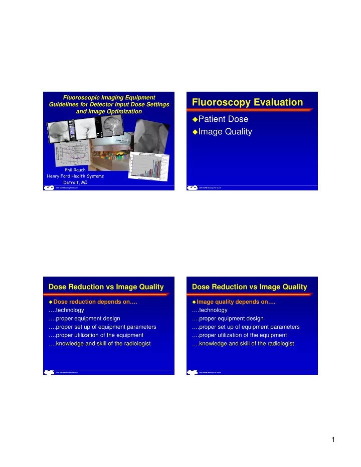
Fluoroscopy Evaluation
Patient Dose Image Quality
2009 AAPM Meeting-Phil Rauch
Dose Reduction vs Image Quality
Dose reduction depends on….
….technology ….proper equipment design ….proper set up of equipment parameters ….proper utilization of the equipment ….knowledge and skill of the radiologist
2009 AAPM Meeting-Phil Rauch
Image quality depends on….