Fish Anatomy & Disease Diagnosis Alex Primus University of - - PowerPoint PPT Presentation
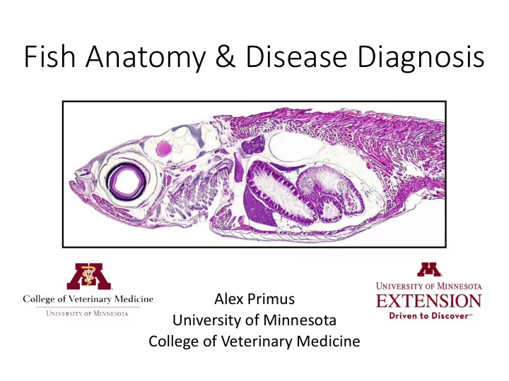
Fish Anatomy & Disease Diagnosis Alex Primus University of Minnesota College of Veterinary Medicine Overview Anatomy Basic Fish Anatomy Gills Diagnostics Basic Advanced State of the Art Dx Why Anatomy &
Overview
- Anatomy
- Basic Fish Anatomy
- Gills
- Diagnostics
- Basic
- Advanced
- State of the Art Dx
Why Anatomy & Diagnostics?
- Anatomy
- The better you know your fish – inside and out – the better
you will be at recognizing disease, managing disease, and keeping your fish healthy
- Recommendation: Take a good look at your fish occasionally
- Get a good sense of what “normal” looks like – inside and out
- Diagnostics
- Some diagnostics can be done on the farm, by the producer
- Help identify disease as early as possible
- Best chance to manage disease early and minimize losses
- Other diagnostics more complex
- The more you know, the better you will be at working with your
vet or diagnostic lab to manage the health of your fish
Basic Fish Anatomy
Basic Perch Anatomy - External
Perch Basic Anatomy
Perch Basic Anatomy
Fish Gills
Gill Health is Extremely Important!
- Involved in:
- Respiration (gas exchange)
- Metabolite excretion (e.g. ammonia)
- Ion exchange (e.g. Na+, Cl-, etc.)
Fish Gills
- Very delicate structures
- Irritants quickly and
significantly decrease function
- Poor Water Quality
- Ectoparasites
- Bacteria
- Chemicals
Protist Parasite Damaged Gill Normal Gill
Disease Diagnosis
Diagnostic Goals
- If fish are sick/dying, identify the cause of that
disease
- Process involves…
- Identification of:
- Gross morphological abnormalities
- Histological abnormalities (at the level of tissues or cells)
- Presence of infectious agents
- Combine Dx results with clinical signs, history, WQ, etc.
- Initiating factors (often poor water quality or stress)
- Factors ultimately resulting in morbidity/mortality
Diagnostic Tools in Fish Health
- Basic
- Gross morphology
- Wet-mounts (skin, fin & gill)
- Advanced
- Bacteriology
- Virology
- PCR
- Histopathology
- State of the Art
- Electron Microscopy
- Whole Genome Sequencing
Gross Morphology
- External signs of Dz
Wet-Mounts
- To evaluate for ectoparasites, external bacterial
infections, and external fungal/saprolegina infections
- Tissues/samples typically evaluated
- Gill clip
- Fin clip
- Skin scrape
Wet-Mounts: Common Pathogens
Trichodina Monogenean Flatworms Saprolegnia Columnaris
Gross Morphology
- Internal signs of Dz
Bacteriology
- Typically to test for systemic bacterial infections
- Use swab to sample sites of interest → inoculate
culture media → incubate
- Ideal sites: Anterior kidney & Brain (sterile sampling)
- Basic media: TSA, BHI, Blood agar
- Other media required for some pathogens
Virology
- Virus Isolation is the gold standard
- Involves inoculating cell culture with tissues of interest
- If virus present → virus infects cells → CPE
- Several cell culture types & temperatures used
- Sensitivity for particular virus dependent on cell type and
incubation temperature
- If CPE, identification of virus requires additional testing
Healthy Cells CPE
Histopathology
- Analysis of tissues on the microscopic level
- Can be used to diagnose a number of diseases
- Involves preserving tissues in fixative → embedding
in solid paraffin block → slicing in very thin sections → staining sections → microscopic analysis
PCR – Polymerase Chain Reaction
- Molecular assay that indicates the presence or
absence of DNA specific to certain pathogens
- Works by amplifying target DNA sequence if present
- Quick, specific, can be very useful (particularly for
viral or bacterial pathogens)
State of the Art Diagnostics
Electron Microscopy
- Uses a beam of electrons to
create an image of specimen
- Much higher magnifications
than a light microscope Whole Genome Sequencing
- Process of determining the
complete DNA sequence of an organism's genome at a single time
(Photo by A. Armien, U. of Minnesota)