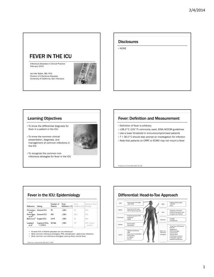
FEVER IN THE ICU Infectious Diseases in Clinical Practice February - - PDF document

fever in the icu
SMART_READER_LITE
LIVE PREVIEW

FEVER IN THE ICU Infectious Diseases in Clinical Practice February - - PDF document

2/4/2014 Disclosures NONE FEVER IN THE ICU Infectious Diseases in Clinical Practice February 2014 Jennifer Babik, MD, PhD Division of Infectious Diseases University of California, San Francisco Learning Objectives Fever: Definition and


slide-1
SLIDE 1

2/4/2014 1

FEVER IN THE ICU

Infectious Diseases in Clinical Practice February 2014 Jennifer Babik, MD, PhD Division of Infectious Diseases University of California, San Francisco

Disclosures

  • NONE

Learning Objectives

  • To know the differential diagnosis for

fever in a patient in the ICU

  • To know the common clinical

presentation, diagnosis, and management of common infections in the ICU

  • To recognize the common non-

infectious etiologies for fever in the ICU

Fever: Definition and Measurement

  • Definition of fever is arbitrary
  • ≥38.3°C (101°F) commonly used, IDSA/ACCCM guidelines
  • Use a lower threshold in immunocompromised patients
  • T < 36.0°C should also prompt an investigation for infection
  • Note that patients on CRRT or ECMO may not mount a fever

O’Grady et al, Crit Care Med 2008, 35:1330.

Fever in the ICU: Epidemiology

Niven et al, J Intensive Care Med 2012, 27:290.

  • At least 50% of febrile episodes are non-infectious!
  • Most common infectious etiologies: PNA, bloodstream, abdominal infections
  • Most common non-infectious etiologies: post-op fever, central fever

Differential: Head-to-Toe Approach

  • Nosocomial meningitis

(post-NSG)

  • Nosocomial meningitis

(post-NSG) CNS

  • Nosocomial Sinusitis
  • URI (hospital-acquired)
  • Nosocomial Sinusitis
  • URI (hospital-acquired)

HEENT

  • Hospital-acquired PNA
  • Empyema
  • ARDS
  • Hospital-acquired PNA
  • Empyema
  • ARDS

Pulmonary

  • Endocarditis
  • Pericarditis
  • Endocarditis
  • Pericarditis

Cardiac

  • Abdominal abscess
  • SBP
  • Acalculous cholecystitis
  • Pancreatitis
  • C. difficile
  • CA-UTI
  • Abdominal abscess
  • SBP
  • Acalculous cholecystitis
  • Pancreatitis
  • C. difficile
  • CA-UTI

GI/GU

  • Osteomyelitis/septic

arthritis

  • Gout
  • Osteomyelitis/septic

arthritis

  • Gout

MSK

  • Cellulitis at PIV/CVC
  • Infected decubitus ulcer
  • Surgical site infection
  • Cellulitis at PIV/CVC
  • Infected decubitus ulcer
  • Surgical site infection

Skin

  • CLABSI
  • Candidemia
  • CLABSI
  • Candidemia

Systemic

  • Drug Fever/Withdrawal
  • DVT/PE
  • Malignancy
  • Rheumatologic
  • Central fever
  • Post-op fever
  • Transfusion reaction
  • Transplant rejection
  • Adrenal insufficiency
  • Drug Fever/Withdrawal
  • DVT/PE
  • Malignancy
  • Rheumatologic
  • Central fever
  • Post-op fever
  • Transfusion reaction
  • Transplant rejection
  • Adrenal insufficiency

Other non- infectious etiologies

slide-2
SLIDE 2

2/4/2014 2 Differential: Head-to-Toe Approach

  • Nosocomial meningitis

(post-NSG)

  • Nosocomial meningitis

(post-NSG) CNS

  • Nosocomial Sinusitis
  • URI (hospital-acquired)
  • Nosocomial Sinusitis
  • URI (hospital-acquired)

HEENT

  • Hospital-acquired PNA
  • Empyema
  • ARDS
  • Hospital-acquired PNA
  • Empyema
  • ARDS

Pulmonary

  • Endocarditis
  • Pericarditis
  • Endocarditis
  • Pericarditis

Cardiac

  • Abdominal abscess
  • SBP
  • Acalculous cholecystitis
  • Pancreatitis
  • C. difficile
  • CA-UTI
  • Abdominal abscess
  • SBP
  • Acalculous cholecystitis
  • Pancreatitis
  • C. difficile
  • CA-UTI

GI/GU

  • Osteomyelitis/septic

arthritis

  • Gout
  • Osteomyelitis/septic

arthritis

  • Gout

MSK

  • Cellulitis at PIV/CVC
  • Infected decubitus ulcer
  • Surgical site infection
  • Cellulitis at PIV/CVC
  • Infected decubitus ulcer
  • Surgical site infection

Skin

  • CLABSI
  • Candidemia
  • CLABSI
  • Candidemia

Systemic

  • Drug Fever/Withdrawal
  • DVT/PE
  • Malignancy
  • Rheumatologic
  • Central fever
  • Post-op fever
  • Transfusion reaction
  • Transplant rejection
  • Adrenal insufficiency
  • Drug Fever/Withdrawal
  • DVT/PE
  • Malignancy
  • Rheumatologic
  • Central fever
  • Post-op fever
  • Transfusion reaction
  • Transplant rejection
  • Adrenal insufficiency

Other non- infectious etiologies

Parts of the Exam to Remember

  • Careful neuro exam
  • Careful neuro exam

CNS

  • Look for nasal discharge
  • Look for nasal discharge

HEENT

  • Back and joint exam
  • Back and joint exam

MSK

  • Examine line sites
  • Take off bandages
  • Peri-anal exam
  • Look for (drug) rashes
  • Examine line sites
  • Take off bandages
  • Peri-anal exam
  • Look for (drug) rashes

Skin

Diagnostics

  • Avoid “automatic” order sets for fever (i.e. the “pan-culture”)
  • Expensive
  • Time-consuming
  • Patient discomfort
  • Unnecessary radiation
  • Transfer out of the controlled unit for imaging/procedures
  • With more testing, you will find more colonizers/contaminants
  • Use a rational approach
  • Start with a history and physical
  • Order labs/diagnostics based on clinical suspicion

O’Grady et al, Crit Care Med 2008, 35:1330.

Case #1

35 y/o man with alcoholic cirrhosis is admitted to the ICU with severe influenza A and is intubated and eventually requires

  • ECMO. He was slowly improving but then over the last 2 days

has starting having fevers to 38.3 with increasing O2

  • requirement. He has trouble following commands when

sedation is lifted. Blood and urine cultures are negative. CXR is unchanged. Head CT shows pansinusitis but is otherwise negative.

Your Next Diagnostic Step is:

  • 1. Sinus puncture
  • 2. Lumbar puncture
  • 3. Mini-BAL or endotracheal aspirate

Defining CAP, HAP, VAP, HCAP

Community- acquired pneumonia (CAP)

PNA not acquired in association with a healthcare setting (i.e. not meeting criteria for HAP or HCAP)

IDSA/ATS Guidelines, Am J Resp Crit Care Med 2005.

slide-3
SLIDE 3

2/4/2014 3 Defining CAP, HAP, VAP, HCAP

Community- acquired pneumonia (CAP) Hospital-acquired pneumonia (HAP)

PNA not acquired in association with a healthcare setting (i.e. not meeting criteria for HAP or HCAP) PNA acquired after 48 hours in the hospital and not incubating at the time

  • f admission.

Risk for MDR pathogens when hospitalized for ≥ 5 days.

IDSA/ATS Guidelines, Am J Resp Crit Care Med 2005.

Defining CAP, HAP, VAP, HCAP

Community- acquired pneumonia (CAP) Hospital-acquired pneumonia (HAP) Ventilator-associated pneumonia (VAP)

PNA not acquired in association with a healthcare setting (i.e. not meeting criteria for HAP or HCAP) PNA acquired after 48 hours in the hospital and not incubating at the time

  • f admission.

Risk for MDR pathogens when hospitalized for ≥ 5 days. PNA acquired after 48 hours of intubation. This is a subset of HAP. Risk by days intubated:

  • Days 1-5 (3%/d)
  • Days 5-10 (2%/d)
  • Days > 10 (1 %/d)

IDSA/ATS Guidelines, Am J Resp Crit Care Med 2005.

Defining CAP, HAP, VAP, HCAP

Community- acquired pneumonia (CAP) Hospital-acquired pneumonia (HAP) Ventilator-associated pneumonia (VAP) Healthcare-associated pneumonia (HCAP)

PNA not acquired in association with a healthcare setting (i.e. not meeting criteria for HAP or HCAP) PNA acquired after 48 hours in the hospital and not incubating at the time

  • f admission.

Risk for MDR pathogens when hospitalized for ≥ 5 days. PNA acquired after 48 hours of intubation. This is a subset of HAP. Risk by days intubated:

  • Days 1-5 (3%/d)
  • Days 5-10 (2%/d)
  • Days > 10 (1 %/d)

PNA in a non-hospitalized patient with any one of the following:

  • Hospitalization for ≥2 days

in the last 90 days

  • Residence in a nursing

home or long-term care facility

  • Intravenous antibiotics,

wound care, dialysis, or chemotherapy within the last 30d

  • Family member with an

MDR pathogen

IDSA/ATS Guidelines, Am J Resp Crit Care Med 2005.

  • To characterize the risk of MDR bacteria, and therefore

inform your empiric antibiotic approach

  • Risk of MDR organisms in PNA:
  • HCAP
  • HAP/VAP with ≥ 5 days in the hospital
  • Immunosuppression
  • Abx in last 90 days
  • High frequency of ABx resistance in a specific unit

Why do we care about these distinctions?

IDSA/ATS Guidelines, Am J Resp Crit Care Med 2005.

VAP: Diagnosis

ST STEP 1 1

Clinical suspicion for PNA? (1) New or progressive CXR infiltrate + (2) 2 out of 3 clinical criteria:

  • F > 38˚C
  •  or  WBC
  • Purulent secretions
  • 69% sensitive, 75% specific
  • With ARDS: consider PNA when

have 1 clinical criteria b/c may not see CXR changes

ST STEP 2 2

Lower respiratory tract culture before ABx

  • Quantitative cultures preferred
  • BAL (cutoff 104 or 105) and

mini-BAL (cutoff 103 or 104) both are ~80% sensitive and specific

  • Even 24 hrs of prior ABx can

make a sample negative

IDSA/ATS Guidelines, Am J Resp Crit Care Med 2005. Klompas, JAMA 2007, 297:1583.

VAP: Diagnosis

ST STEP 3 3

  • Start empiric therapy
  • Re-evaluate each day based on

culture results, clinical course

  • Consider stopping ABx if

cultures are negative

  • Other diagnostics:
  • Sensitivity of blood cultures

<25%

  • Thoracentesis if an effusion

is large or the patient is toxic

IDSA/ATS Guidelines, Am J Resp Crit Care Med 2005. Raman, Crit Care Med 2013, 41:0,

slide-4
SLIDE 4

2/4/2014 4

VAP/HAP: Empiric ABx (IDSA Guidelines)

Ceftriaxone

  • r

Fluoroquinolone

  • r

Ertapenem Risk Factors for MDR Pathogens Present?

  • HCAP
  • HAP/VAP with ≥ 5 days in the hospital
  • Immunosuppression
  • Abx in last 90 days
  • High frequency of ABx resistance in a specific unit

No Yes

Linezolid or Vancomycin + Anti-pseudomonal beta lactam + Anti-pseudomonal FG or AG

IDSA/ATS Guidelines, Am J Resp Crit Care Med 2005.

*Use local resistance patterns for guidance

  • Chastre et al: RTC of 400 pts dx’d with VAP by quantitative BAL,

randomized to 8 vs. 15 days of ABx therapy

  • No difference in: mortality, recurrent infections, length of ICU stay
  • 8-day group had:
  • More ABx-free days (9 vs 13%)
  • Less MDR organisms if had recurrent infections (42 vs 65%)
  • But…higher pulmonary reinfection rate (41 vs 25%) if had a glucose

nonfermenter (Pseudomonas, Acinetobacter, or Stenotrophomonas)

  • Take-home:
  • Pseudmonas, Acinetobacter, Stenotrophomonas: 14 (or 15) days
  • MRSA: 7-21 days depending on extent of infection (IDSA MRSA guidelines)
  • 7 (or 8) days for everyone else

Duration of ABx

Chastre, JAMA 2003, 290:2588.

Linezolid vs. Vancomycin for MRSA?

  • Phase 4, double-blind, randomized, controlled multicenter

trial, pts with MRSA HAP/VAP/HCAP

  • IV linezolid 600 mg Q12h vs. IV vancomycin (15 mg/kg Q12h)
  • Vanco dose-optimized by unblinded pharmacist
  • Treated for 7-14 days (up to 21 d if bacteremia)
  • Primary outcome: Clinical response
  • Cure defined as: resolution of clinical signs and sx, improvement or

lack of progression of CXR, no additional abx required

Wunderink et al, Clin Infect Dis 2012; 54: 621.

Linezolid vs. Vancomycin: Outcomes

Wunderink et al, Clin Infect Dis 2012; 54: 621.

Study Conclusions

  • Linezolid has a modest benefit in clinical response over

vancomycin in MRSA HAP/VAP

  • Limitations:
  • ? Vanco pts sicker: Compared to linezolid group, more ventilated,

concurrent MRSA bacteremia, kidney disease and diabetes

  • No difference in hard outcomes:
  • No mortality benefit
  • Did not evaluate length of ICU stay, length of hospitalization, mechanical

ventilation

Wunderink et al, Clin Infect Dis 2012; 54: 621.

Nosocomial Transmission of Resp Viruses

  • Has been described with:
  • RSV
  • Influenza
  • Parainfluenza
  • Adenovirus
  • Transmission can be between

patients or from visitors/staff

Aitken and Jeffries, Clin Micro Rev 2001, 14:528.

slide-5
SLIDE 5

2/4/2014 5 HAP/VAP: Take Home Points

  • Think about risk factors for MDR pathogens and use that to

guide empiric therapy

  • Diagnosis is based on a combination of clinical and

microbiologic paramters

  • Duration of therapy 7 days with the exception of the glucose

nonfermenters +/- MRSA

  • Consider linezolid for MRSA if not responding to vancomycin

Nosocomial Sinusitis

  • Epidemiology:
  • Radiographic sinusitis in 25-75% of ICU pts
  • But etiology of nosocomial fever in ~5%
  • Radiographic sinusitis ≠ infectious sinusitis
  • Micro: Pseudomonas, S. aureus, can be polymicrobial
  • Clinical: classic signs/sx of sinusitis often absent
  • Dx: CT, aspirate by ENT to confirm dx and guide ABx therapy
  • Treatment duration: 7 days

O’Grady et al, Crit Care Med 2008, 35:1330. George et al, Clin Infect Dis 1998, 27:463. Talmor et al, Clin Infect Dis 1997, 25:1441. Borman et al, JAMA 1992, 164:412. Stein and Kaplan, Curr Opin Infect Dis 2005, 18:147.

A 65 y/o M is admitted with an STEMI. 4 days into his hospitalization he spikes a fever to 39, drops his SaO2 to the low 90s on RA, and becomes altered. He has a foley. He is started on vancomycin and pip/tazo and improves. Work-up reveals:

  • CXR with a new LLL infiltrate
  • Blood cultures and sputum culture negative at 48h
  • UA (from his catheter) shows 30 WBC, Urine cx >100K VRE

Case #2 What would you do with his ABx?

  • 1. Continue pip-tazo and d/c vanco
  • 2. Continue pip-tazo and change vancomycin to linezolid to

cover VRE

CA-UTI is a Diagnosis of Exclusion!

  • Catheter-associated bacteriuria is common (up to 25% of

patients with short term catheters)

  • CA-bacteriuria usually represents colonization (asymptomatic

bacteriuria = ASB) and NOT infection

  • What about pyuria?
  • Pyuria is common in catheterized patients (up to 75% with short term

catheters)!

  • The presence or degree of pyuria cannot differentiate ASB from UTI
  • But, the absence of pyuria suggests an alternative diagnosis

Hooton, CID 2010. Nicolle, CID 2005. Tambyah, Arch Intern Med 2000.

  • 1. Patient with a catheter currently or within the last 48 h
  • 2. Symptoms or signs c/w UTI
  • 3. No other source of infection (i.e., diagnosis of exclusion)
  • 4. ≥103 cfu of ≥1 bacterial species in a urine culture

Catheter-associated UTI: Definition

Hooton, CID 2010.

slide-6
SLIDE 6

2/4/2014 6

  • What are “signs and symptoms compatible w/UTI” in a

patient with a catheter?

  • New onset or worsening of fever, rigors, altered mental status, malaise
  • r lethargy with no other identified cause
  • Flank pain or CVAT
  • Acute hematuria
  • Pelvic discomfort
  • Spinal cord injury patients: increased spasticity, autonomic dysreflexia,
  • r sense of unease

The million dollar question…

Hooton, CID 2010.

  • First, STOP: do you need a UA/urine culture if you have a high

suspicion for an alternative source (eg PNA)?

  • Second: if you are sending a urine culture, always get a UA
  • How to get the best culture?
  • A catheter culture may not accurately reflect what is in the bladder
  • If a catheter has been in place for >2 weeks, change it and get a Ucx

from the newly placed catheter

  • In a patient with a condom cath, get a specimen from a freshly placed

condom cath after cleaning the glans because skin can be colonized

Diagnosis

Hooton, CID 2010.

Sign Signs and and Sym Symptoms of Syst

  • f Systemic Inf

Infection n withou thout classic UT lassic UTI s signs/ s s/ sx

Alternate Diagnosis Likely? (Signs/ sx of other illness present) Yes Do not order U/A, urine cx No Send U/A, urine cx U/A, urine cx (-) Do not treat for UTI U/A (-), urine cx (+) Asymptomatic bacteriuria

  • Do not Rx for UTI*
  • Look for alternate dx

U/A (+), urine cx (+) Treat for UTI

  • If no alternate dx

identified *Exceptions: pregnancy, pre-urological procedure, neutropenic host

U/A (+), urine cx (-) On abx No abx Do not Rx for UTI

Empiric Therapy for Complicated UTI

  • Community acquired:
  • Ertapenem
  • Ceftriaxone
  • Healthcare associated:
  • Pip/tazo
  • Ertapenem
  • Ceftriaxone
  • Duration:
  • 7 days if there is prompt resolution of symptoms
  • 10-14 days if response is delayed
  • Catheter change?
  • Yes, if the catheter has been in for >2 weeks, change it
  • This has been associated with:
  •  CA-bacteriuria and CA-UTI at 28d
  •  time to resolution of sx

Treatment

Hooton, CID 2010.

  • Asymptomatic candiduria:
  • In general, don’t treat!
  • Change the foley: can eliminate candiduria in 20-40%
  • Exceptions: Patients at high risk of dissemination
  • Neutropenia
  • Patients undergoing urologic procedures
  • Symptomatic candiduria: treat

Candiduria: Who to Treat?

Pappas, CID 2009.

slide-7
SLIDE 7

2/4/2014 7

1st line: Fluconazole

  • Excellent urine levels, 10-fold higher than serum levels
  • Can get concentrations in the urine that are higher than the

MIC for organisms that are intermediate or resistant

  • 200-400mg PO daily

Candida UTI: Treatment Options

Fisher, CID 2011.

  • Can try fluconazole and re-check Ucx (if not systemically ill)
  • Other options:
  • Flucytosine
  • Amphotericin B
  • Ampho bladder washes: Resolve candiduria in >90% but  relapses
  • Other azoles?
  • Vori, posa, itra have poor urinary penetration
  • Caspofungin?
  • Poor urinary penetration, but use if suspect systemic disease

Fluconazole-Resistant Candida UTI

Fisher, CID 2011.

CA-UTI: Take-home points

  • ASB and pyuria are common in patients with a foley
  • To diagnose a CA-UTI, the patient must have:
  • Signs and symptoms compatible with UTI
  • No other source for infection
  • Treat for 7-14 days depending on clinical response
  • Candiduria is almost always asymptomatic and does not

require treatment

Case #3

A 55 year old man is admitted to the ICU with severe gallstone

  • pancreatitis. He has been afebrile and slowly improving on

TPN and with conservative measures. He spikes a fever to 39 associated with new hypotension and rigors. Blood cultures drawn from his triple lumen subclavian line turn positive 6 hours before his peripheral blood culture. Both are growing E. coli.

The most sensitive test to determine the source of the E coli is:

  • 1. Differential time to positivity
  • 2. Inflammation around the central line exit site
  • 3. Remove the catheter and culture the tip

The IDSA Guidelines recommend:

  • 1. Pull the line
  • 2. Ok to attempt line salvage with ABx lock therapy
  • 3. Ok to attempt line salvage by giving systemic ABx through

the line

slide-8
SLIDE 8

2/4/2014 8 CLABSI: Diagnosis

  • Clinical findings unreliable:
  • Inflammation at the exit site is extremely insensitive (<3%)
  • Positive peripheral bcx and > 15 CFU/plate of same organism

from catheter tip

  • 79% sensitive, 92% specific
  • But >80% of catheters withdrawn b/c of clinical suspicion of CLABSI are

removed unnecessarily

  • Quantitative blood cultures (line vs peripheral) may be most

sensitive/specific, but not routinely available

Mermel et al CID 2009. Safdar and Maki, Crit Care Med 2002, 30:2632.

CLABSI: Differential Time to Positivity

  • Allows for diagnosis without removing the line
  • Draw culture from central line and peripheral blood at the

same time

  • CLABSI = blood culture drawn from central line turns positive

at least 2 hrs before peripheral culture turns positive

  • Test characteristics
  • 95% sensitive
  • 90% specific (but only ~40% specific for yeast)

Liñares, Clin Infect Dis 2007, 44:827. Bouza et al, Clin Infect Dis 2007, 44:820. Bouza et al, Clin Microbiol Infect 2013, 19: E129.

CLABSI: Management

  • Possible scenarios:
  • Line (+)/ peripheral (+)
  • DTTP ≥ 2 hrs  CLABSI  remove line and treat
  • DTTP < 2 hrs  look for another source and treat
  • Line (+)/ peripheral (-)  colonization/ contaminant
  • Duration of treatment (with line removal):
  • 5-7 days: coagulase negative Staphylococci
  • 7-14 days: GNRs, Enterococcus
  • 14 days: S. aureus, Candida

Mermel et al CID 2009

CLABSI: Line Management

  • Remove line whenever possible:
  • Guidewire exchange only if no alternative insertion site
  • If septic, hemodynamically unstable, persistent bacteremia  remove line
  • Long term catheters: remove if S aureus, Pseudomonas, Candida
  • Short term catheters: remove if S aureus, GNRs, Candida, Enterococcus
  • Line salvage
  • Antibiotic lock therapy + systemic antibiotic therapy
  • Studied primarily in long-term catheters

Mermel et al CID 2009

Case #4

85 y/o man is admitted with fever and respiratory failure to the ICU and treated with vanc/pip-tazo. He initially responds but then 5 days into therapy he began spiking high fevers up to 39˚C daily. His respiratory status is unchanged. He is escalated to vanc/meropenem with no change in his fever or respiratory status after another 5 days. Extensive work-up for

  • ther sources of infection is negative.

What is Your Next Step?

  • 1. Change vanco to linezolid
  • 2. Add tobramycin
  • 3. Stop antibiotics
slide-9
SLIDE 9

2/4/2014 9 Drug Fever

  • 3-4 % of all drug reactions
  • Multiple mechanisms:
  • Altered Thermoregulatory Mechanisms (eg amphetamine)
  • Drug Administration (eg amphotericin)
  • Pharmacologic Effects (eg Jarisch-Herxheimer Reaction)
  • Idiosyncratic Reactions (eg malignant hyperthermia)
  • Immune-Mediated/Hypersensitivity Reactions (eg most ABx)

Drugs Associated with Drug Fever

Patel, et al. Pharmacotherapy 2010; 30(1):57-69.

Clinical

  • May appear well and be unaware of fevers (but not

necessarily)

  • No typical fever pattern
  • Pulse-temperature dissociation (11%)
  • Expected pulse = [(last digit in F -1 ) x 10] +100
  • Rash (5-10%)
  • Eosinophilia (~20%)
  • Labor. Drug Intell Clin Pharm 1986; 20:413-20. Mackowiak, Ann Int Med 1987; 106:728-33.

Mackowiak, et al. Ann Int Med 1987; 106:728-33. Foster, et al. Med Clin North Am 1966;42:523-39

Class of Offending Agent Episodes Lag Time Mean Median SD N

  • -------------------Days--------------------

Cardiac 36 44.7 10 131.1 Antimicrobial 44 7.8 6 8.4 Antineoplastic 11 6 0.5 12.3 CNS 24 18.5 16 15.4 Other 20 18.8 7 34.1

  • But with re-challenge, fever can occur within hours

Timeline of Fever Onset Fever Characteristics

  • Fever is high
  • Usually defervesce within

1-2 d of stopping the drug

Mackowiak, et al, Ann Intern Med 1987, 106:728.

38.5 39 39.5 40 40.5 41 41.5 0.5 1 1.5 2 Temp (˚C) Days to defervescence

Treatment

  • Discontinue all potentially causative meds, together or sequentially
  • In cases where benefit > risk in continuing, can try to pre-treat:
  • Corticosteroids
  • Antihistamines
  • But watch for S/Sx of progression of hypersensitivity
  • Rechallenge will usually cause recurrence of fever within a few

hours, confirming the diagnosis

  • If fever was accompanied by severe adverse effects, avoid rechallenge
  • Important to document suspected drug fever in the allergy section with as

much detail of associated symptoms as possible

Patel, et al, Pharmacotherapy 2010, 30:57.

slide-10
SLIDE 10

2/4/2014 10 Cross-Reactivity of Antibiotics?

 Change to another class if possible (i.e. Beta-lactam to

fluoroquinolone

 No studies exist which address drug fever cross reactivity

specifically – focus is on all symptoms of hypersensitivity

Joint Task Force on Practice Parameters. Ann Allergy Asthma Immunol 1999; 83:665-700.

Drug Fever: Take Home Points

  • Always consider it in the ddx for fever in the hospital
  • Look for eos, temp-pulse dissociation, rash although

remember these are present in <20% of cases

  • Consider stopping the ABx or swtiching classes if you really

suspect it

  • Remember to document drug fever as an allergy!

VTE and Fever

  • Seen in 5-15% of

patients presenting with PE/DVT

  • Characteristics:
  • Usually <38.3
  • Peaks on day of PE
  • Gradually subsides

within 1 week

10 20 30 Distribut ribution o

  • f F

Fever a r and PE ( (Pioped) ped)

Stein et al, Chest 2000, 117:39. Nucifora et al, Circulation 2007, 115:e173. Barba et al, J Thromb Thrombolysis 2011, 32:288.

# patients

Central Fever

  • Accounts for ~50% of fever in the

Neuro-ICU

  • Seen in patients with brain tumors,

SAH, intraventricular hemorrhage

  • Associated with vasospasm
  • Appears within 72 hours of admission,

persists for longer than infectious causes of fever

  • No difference in height of fever

Hocker et al, JAMA Neurol 2013, 70:1499.

Can ARDS Itself Cause Fever?

  • The fibroproliferative phase of ARDS

can cause fever and leukocytosis that is indistinguishable form infection

  • Open lung biopsy in 7/9 patients with

late ARDS found fibroproliferative phase of diffuse alveolar damage and no evidence of infection

  • So…probably, but would look very hard

elsewhere and this is a diagnosis of exclusion

Meduri et al, Chest 1991, 100:943.

65 y/o F in the ICU for a prolonged course after a Whipple

  • procedure. Her course has included a VAP and UTI and she

has received multiple courses of antibiotics. She has been spiking fevers for the last 3 days despite linezolid and

  • meropenem. You get a call from the micro lab that 1/2 blood

cultures (peripheral) is growing yeast.

Case #5

slide-11
SLIDE 11

2/4/2014 11 The most appropriate next step is:

  • 1. Start voriconazole
  • 2. Start fluconazole
  • 3. Start caspofungin
  • Yeast in the blood almost always = candida
  • Rarely it could be cryptococcus in the right host (e.g., HIV,

transplant)

What is the ddx for “yeast in the blood”?

  • So you know it’s candidemia, so now what do you do?
  • You need to do 3 things:

1.

Start caspofungin

2.

Evaluate for source  pull lines

3.

Eye exam

Back to the case…

  • What kinds of candida are there?
  • C albicans (50-65%)
  • C glabrata (~20%) – can be fluconazole resistant
  • C parapsilosis (6-17%)
  • C tropicalis (7-11%)
  • C krusei (2%)
  • C lusitaniae (<1%)
  • C dublinensis (<1%)

Why caspofungin and not fluconazole?

Messer, J Clin Micro 2003. Pfaller, J Clin Micro 2010.

  • Dosing:
  • 70mg IV x 1 then 50mg IV daily
  • No adjustment needed for renal failure or HD/CVVH
  • SEVERE hepatic disease: decrease maintenance dose to 35mg qday

(*based on PK study of drug levels, not adverse effects)

  • Drug-drug interactions:
  • Increase maintenance dose to 70mg when given with phenytoin,

rifampin, carbamezapine, dexamethasone, nevirapine, or efavirenz

  • Adverse effects: very well tolerated, can get elevation of LFTs

Caspofungin Basics

Mistry et al, J Clin Pharmacol 2007.

  • The candida comes back as C glabrata…anything else to do?
  • Yes – ask the lab for fluconazole susceptibilities!
  • If it comes back sensitive to fluconazole  switch to

fluconazole to finish the course.

Back to the Case…

slide-12
SLIDE 12

2/4/2014 12 Candida Susceptibilities

  • C albicans or C tropicalis:
  • Fluc resistance very rare: C albicans ~1-2 %, C tropicalis ~4%
  • Fluconazole is drug of choice

Pappas, CID 2009. Pfaller, JCM 2010.

  • C parapsilosis:
  • Caspo MICs are in general higher, although clinical significance unclear
  • Fluconazole is drug of choice (~4% fluc resistance seen)

Candida Susceptibilities

Pappas, CID 2009. Pfaller, JCM 2010. Kale-Pradhan, Pharmacotherapy 2010.

  • C glabrata:
  • Fluc resistance is ~15% nationally, vori resistance ~10%
  • Caspo is drug of choice to start, then narrow to fluc based on sensitivities (or

consider vori as oral step-down alternative if sensitive)

Candida Susceptibilities

Pappas, CID 2009. Pfaller, JCM 2010.

  • C krusei:
  • Intrinsic fluconazole resistance
  • Caspo is drug of choice, consider step-down to vori as oral option

Candida Susceptibilities

Pappas, CID 2009.

  • C lusitaniae:
  • Can be amphotericin resistant

Candida Susceptibilities

Pappas, CID 2009.

IDSA guidelines:

  • If no metastatic foci of infection, treat for 2 weeks from date
  • f 1st negative culture (so be sure to get surveillance cx)
  • This is based on the results of several prospective,

randomized trials in which treatment for 2 weeks was associated with few complications and relapses

Duration of Therapy

Pappas, CID 2009.

slide-13
SLIDE 13

2/4/2014 13

  • Remove cathethers if possible:
  • Often difficult to tell in an individual patient if catheter or GI is the

source

  • Exception: C parapsilosis is often catheter-associated
  • Removal is associated with more rapid clearance of blood cultures and

decreased mortality

  • Note that this data is less compelling in neutropenic patients, so

recommendation to remove catheters is less strong in this population

Pull the line!

  • Rule out chorioretinitis (seen in ~10%) or endophthalmitis

(seen in 1-2%)

  • This is not an emergency (unless having visual symptoms)
  • In fact, may increase your sensitivity by waiting ~1 week after

starting therapy

Get an Eye Exam

Oude Lashof , CID 2011.

  • Intravitreal injections (ampho)
  • Longer duration of therapy (4-6 weeks)
  • Choose an agent with good eye penetration
  • Azoles (voriconazole>fluconazole)
  • Ampho + 5-FC
  • NOT echinocandins (have poor ocular penetration)

Why does this matter?

  • 1. Start an echinocandin empirically
  • Check surveillance cx in 48hr
  • Get susceptibilities if it’s C glabrata and change to fluc if sensitive
  • Change to fluc if it’s a susceptible species (albicans, tropicalis,

parapsilosis)

  • Treat for 2 weeks from the date of the 1st negative culture
  • 2. Pull the line
  • 3. Eye exam
  • If positive, use vori if sensitive
  • Duration of therapy 4-6 weeks for eye involvement

Candidemia: Take Home Points

What is the role of procalcitonin in the ICU?

  • Diagnosis of sepsis vs. non-infectious SIRS
  • Meta-analysis of 18 studies suggests low diagnostic accuracy (sensitivity/

specificity of ~ 70%)

  • Aid in antibiotic discontinuation
  • Meta-analysis of 14 RCTs investigating PCT algorithms for antibiotic

therapy decisions

  • Reduced antibiotic exposure by 20-37%
  • No mortality difference
  • Decr LOS in some studies, not in others

Tang et al Lancet ID 2007; Schuetz Arch Intern Med 2011.

Fever in the ICU: Fever of Too Many Origins?

Horowitz, NEJM 2013, 368:3.

slide-14
SLIDE 14

2/4/2014 14 Thank you!