
Thermo Fisher Scientific • Street Address • City, ST ZIP Code • thermofisher.com
Abhay Kotecha, Bart Buijsse, Michael Janus, Lingbo Yu, Hans Raaijmakers
Fast structure determination for proteins and small molecules – a MicroED solution
ABSTRACT
In this session, you will learn how micro electron diffraction (MicroED) allows fast, high-resolution 3D structure determination
- f small chemical compounds and biological macromolecules. To
efficiently collect diffraction datasets of nano-crystals, a cryo-TEM is equipped with a specially designed diffraction camera (Ceta-D) and a MicroED package. The latter combines the necessary hardware components, as well as optimized optical settings and specialized EPU-D Software for automated data collection. Combined with the intrinsic microscope performance, the data collection is fully automated and can be realized in a matter of minutes.
INTRODUCTION
MicroED is a new technique for structure determination of biological macromolecules and small molecules:
CONCLUSIONS
- Fast
atomic-resolution 3D structural information. Diffraction data from nanocrystals in minutes.
- Instant productivity. Nanocrystals as small as 100 nm can
be readily analyzed, removing the burden of growing large crystals (as with X-ray crystallography). Also reduces the amount of sample material required. Mixtures of different polymorphs and compounds can be analyzed.
- Complete turnkey solution. Including hardware, software
and support from one single vendor. Acquired data can be readily processed using established reconstruction packages for X-ray crystallography.
- 2-in-1 solution. MicroED and single particle analysis (SPA)
can be performed on the same cryo-electron microscope. This solution is compatible with new microscopes but is also retrofittable on existing units.
FURTHER INFORMATION
Please scan this QR-code to get to the website containing the MicroED introduction video as well as a download link for the PDF describing the MicroED package and the Ceta-D camera.
EXAMPLES
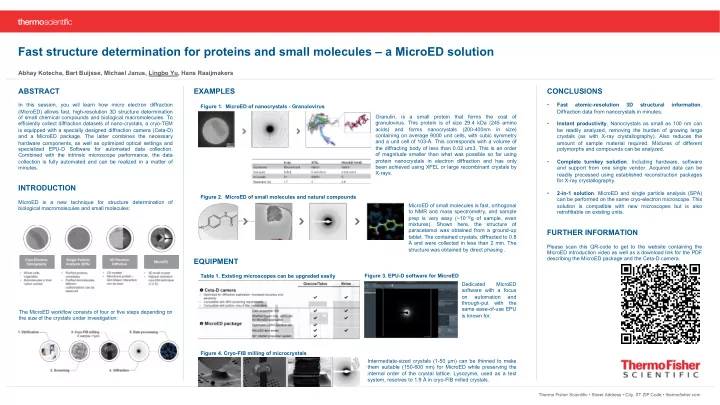
Granulin, is a small protein that forms the coat
- f
- granulovirus. This protein is of size 29.4 kDa (245 amino
acids) and forms nanocrystals (200-400nm in size) containing on average 9000 unit cells, with cubic symmetry and a unit cell of 103-Å. This corresponds with a volume of the diffracting body of less than 0.02 um3. This is an order
- f magnitude smaller than what was possible so far using
protein nanocrystals in electron diffraction and has only been achieved using XFEL or large recombinant crystals by X-rays. The MicroED workflow consists of four or five steps depending on the size of the crystals under investigation: Figure 1. MicroED of nanocrystals - Granulovirus Figure 2. MicroED of small molecules and natural compounds MicroED of small molecules is fast, orthogonal to NMR and mass spectrometry, and sample prep is very easy (~10-12g of sample, even mixtures). Shown here, the structure
- f
paracetamol was obtained from a ground-up
- tablet. The contained crystals, diffracted to 0.8
Å and were collected in less than 2 min. The structure was obtained by direct phasing .
EQUIPMENT
Table 1. Existing microscopes can be upgraded easily Figure 3. EPU-D software for MicroED Dedicated MicroED software with a focus
- n
automation and through-put with the same ease-of-use EPU is known for. Figure 4. Cryo-FIB milling of microcrystals Intermediate-sized crystals (1-50 µm) can be thinned to make them suitable (150-600 nm) for MicroED while preserving the internal order of the crystal lattice. Lysozyme, used as a test system, resolves to 1.9 Å in cryo-FIB milled crystals.