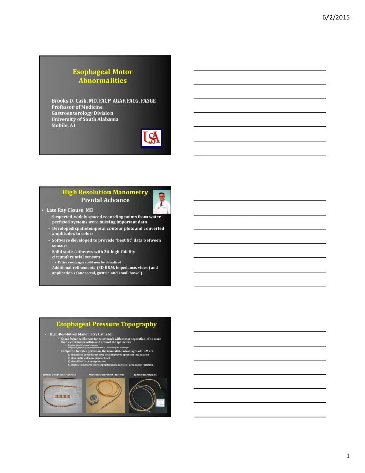

6/2/2015 Esophageal Motor Abnormalities Brooks D. Cash, MD, FACP, AGAF, FACG, FASGE Professor of Medicine Gastroenterology Division University of South Alabama Mobile, AL High Resolution Manometry Pivotal Advance • Late Ray Clouse, MD – Suspected widely spaced recording points from water perfused systems were missing important data – Developed spatiotemporal contour plots and converted amplitudes to colors – Software developed to provide “best fit” data between sensors – Solid state catheters with 36 high ‐ fidelity circumferential sensors • Entire esophagus could now be visualized – Additional refinements (3D HRM, impedance, video) and applications (anorectal, gastric and small bowel) Esophageal Pressure Topography • High ‐ Resolution Manometry Catheter • Spans from the pharynx to the stomach with sensor separation of no more than a centimeter within and around the sphincters. – Greater than 32 pressure sensors – Temporal frequency response matched to the zone of the esophagus • Compared to water perfusion, the immediate advantages of HRM are: 1) simplified procedural set up with improved sphincter localization – 2) elimination of movement artifact – 3) simplified data interpretation – 4) ability to perform more sophisticated analysis of esophageal function. – Sierra Scientific Instruments Medical Measurement Systems Sandhill Scientific Inc. Each sensor has 12 pressure sensitive segments 1
6/2/2015 Functional Imaging of Esophageal Peristalsis HIGH ‐ RESOLUTION MANOMETRY 40 mmHg 0 Manometric port 0 2.5 4.5 5.6 7.1 8.5 8.8 11.0 13.0 Clouse Plot True Functional Imaging of Esophageal Peristalsis ESOPHAGEAL PRESSURE TOPOGRAPHY Pressure mmHg ≥ 110 Clouse Plot 90 70 50 30 Manometric port 10 ‐ 10 0 2.5 4.5 5.6 7.1 8.5 8.8 11.0 13.0 NU IRB 2
6/2/2015 Pressure Topography of Esophageal Motility: What does it add? • More akin to an imaging modality – Defines important anatomical landmarks and abnormalities – Refines measurement of important motor events • EGJ relaxation • Peristaltic timing velocity • Contractile activity/force/amplitude – Defines intra ‐ luminal pressurization patterns – Permits pattern recognition 3 Main Steps in Diagnostic Approach to a High Resolution Manometry Test 1. Assess EGJ anatomy and function 2. Assess esophageal body function 3. Review pressurization patterns These 3 steps will permit diagnosis of most esophageal motor abnormalities * Some changes in prioritization with recent Chicago Classification update (v3.0) Anatomy of a High Resolution Esophageal Manometry Test 3
6/2/2015 STEP 1| Assess the EGJ Anatomy and Function • Determine if hiatus hernia is present • Confirm that the catheter has crossed the EGJ and diaphragm DEEP BREATH Integrated relaxation pressure: The IRP will determine whether outflow obstruction at the EGJ is evident. Disorders are separated at this point, determined by those with or without outflow obstruction at the EGJ. Integrated Relaxation Pressure (IRP) : Mean EGJ pressure measured with a sleeve for 4 contiguous or non‐contiguous seconds of relaxation in the 10‐second window following deglutitive UES relaxation. • The upper limit of normal using ManoScan is 15 mmHg. IRP INTEGRATED RELAXATION PRESSURE IRP INTEGRATED RELAXATION PRESSURE 3 Assess the EGJ Anatomy and Function 4
6/2/2015 STEP 2 | Assess Esophageal Body Function • Peristaltic integrity: either intact, weak or failed • Contractile deceleration point (CDP): anatomic separation point (between tubular esophagus and phrenic ampulla) • Distal Latency (DL): timing of esophageal peristalsis • will define the swallow as premature or normal latency • Distal contractile index (DCI): vigor of the distal esophageal contraction • Contractile front velocity (CFV): speed of esophageal contractions • previously used to define rapid contraction • no longer considered meaningful Peristaltic Breaks: Gaps in the 20 mmHg isobaric contour of the peristaltic contraction between the UES and EGJ, measured in axial length. LARGE BREAK FAILED SWALLOW Contractile Deceleration Point (CDP): The inflection point along the 30 mmHg isobaric contour where propagation velocity slows, demarcating the tubular esophagus from the phrenic ampulla. CDP CONTRACTION DECELERATION POINT (2) Distal Latency (DL): Interval between UES (1) relaxation and the CDP (2), expressed in seconds. Normal DL is >4.5 sec. UES RELAXATION (1) CDP CONTRACTION DECELERATION POINT (2) DL DISTAL LATENCY DL: 7.8 sec CDP CONTRACTION DECELERATION POINT (2) 5
6/2/2015 STEP 3 | Pressurization Patterns Each swallow should be evaluated using the IBC tool to document an isobaric pressurization above 30 mmHg. Achalasia: HRM led to the identification of three discernible achalasia types. Each subgroup represents a distinct clinical entity, each with significantly different biomechanics and treatment outcomes. Type I patients do significantly better with Heller myotomy than with pneumatic dilatation, and Type III patients exhibit the worst prognostic outcome. Achalasia TYPE I Failed peristalsis with abnormal IRP - no esophageal function There is no significant pressurization within the body of the esophagus. Therefore, this would be classified as failed peristalsis with abnormal IRP . In the absence of esophageal body contractility, the IRP threshold of >10 mmHg is used to distinguish Type I Achalasia from absent peristalsis. 5 Major Disorders of Esophageal Peristalsis • Achalasia • Hypertensive LES/EGJ Outflow obstruction • (Nutcracker esophagus) • Jackhammer esophagus • Distal esophageal spasm (DES) • Absent peristalsis Pressure Topography of Esophageal Motility The Chicago Classification Neurogastroenterology and Motility, 2015;27;160 ‐ 74. 6
6/2/2015 Chicago Classification 3.0 Changes • Use median rather than mean cutoff value for IRP • Use lower IRP cutoff for type I achalasia (platform specific) • Panesophageal pressurization with ≥ 20% swallows with 100% failed contractions is type II achalasia irrespective of IRP • Emphasize heterogeneity of conditions potentially causing EGJ outflow obstruction • Modify hypercontractile esophagus to ≥ 20% swallows with DCI >8000 mmHg x s x cm • Substitute ‘absent contractility’ for ‘aperistalsis’ or ‘absent peristalsis’ to differentiate from other scenarios where peristalsis is absent (e.g., achalasia) Chicago Classification 3.0 Changes • Rename ‘minor disorders of peristalsis’ • Eliminate small breaks (2–5 cm) in the 20 ‐ mmHg isobaric contour as a criterion of abnormality • Eliminate rapid CFV (>9 cm/s) as a criterion of abnormality • Eliminate the designation of ‘hypertensive peristalsis’ (DCI 5000–8000 mmHg x s x cm) (no more Nutcracker) • Adopt the ‘ineffective esophageal motility’ (IEM) designation from conventional manometry • Eliminate ‘frequent failed peristalsis’ as a distinct diagnostic entity • Incorporate new data from studies of multiple repetitive swallows into the criteria for IEM Disorders with EGJ Outflow Obstruction The Chicago Classification A c halasia IRP ≥ ULN AND • Subtype I: No contractility 100% failed peristalsis or Subtype II: ≥ 20% PEP • Y es spasm • Subtype III: ≥ 20% spasm (DL<4.5s) No IRP ≥ upper limit of normal EGJ Outflow Obstruction AND Incompletely expressed • some instances of intact or Y es achalasia weak peristalsis • Mechanical obstruction Neurogastroenterology and Motility, 2015;27;160 ‐ 74. 7
6/2/2015 Achalasia • Dysphagia, wt loss, regurgitation, halitosis, GERD sxs • Immune ‐ mediated disease targeting esophageal myenteric plexus (neurons and ganglia) – Antineuronal Abs, inflammatory cells, cytokines, immunoglobulins, complement – Achalasia subtypes may represent differential degree of immune activation/selectivity (cell vs humoral) – HSV ‐ 1 implicated as trigger Kahrilas PJ, et al. Gastroenterology 2013;145:954‐66. High ‐ Resolution Manometry: Achalasia subtypes Type III Type I Type II mmHg 150 air 100 liquid 50 30 IRP= 22.3 mmHg IRP= 24.2 mmHg IRP= 29.8 mmHg 0 5 seconds 5 seconds 5 seconds air diverticulum EGJ EGJ EGJ contraction Clinical Evolution of Achalasia Assessing clinically relevant phenotypes Early Chronic Late Type II or III Type II/III ‐‐ I Type I NU IRB 8
6/2/2015 Achalasia Mimics • Malignancy (Pseudoachalasia) • Chaga’s disease • Amyloidosis • Postvagotomy • Neurofibromatosis • Sarcoidosis • MEN IIb Hypertensive LES • Presentation: Chest pain/dysphagia/globus – May be an achalasia variant • Dx: LES pressure > 35 mmHg AND failure to relax below IRP of 15 mmHg – Normal peristalsis • More important than pressures: failure of full relaxation at LES – Incomplete bolus transfer • Can overlap with other spastic esophageal conditions – May need additional provocation (bread swallow, multiple rapid swallows, solid swallows) – EUS recommended prior to therapy to exclude infiltrative or compressive disease (eg malignancy) EGJ Outflow Obstruction A:EGJOO:achalasia phenotype B:EGJOO: Mechanical mmHg 150 Normal peristalsis 100 Locus of Compartmentalized diverticulum pressurization above EGJ 50 30 IRP= 27.2 mmHg IRP= 22.3 mmHg 0 Barium tablet localized 12 mm restriction Large diverticulum 4 cm above EGJ EGJ 9
Recommend
More recommend