Duplex Ultrasound Evaluation Duplex Ultrasound Evaluation Reversed - - PowerPoint PPT Presentation

duplex ultrasound evaluation duplex ultrasound evaluation
SMART_READER_LITE
LIVE PREVIEW

Duplex Ultrasound Evaluation Duplex Ultrasound Evaluation Reversed - - PowerPoint PPT Presentation

4/14/2016 Disclosures: Speaker honorarium for W.L. Gore When Do You Need to Treat and Associates, 9/2015. Venous Perforators?.. and How to Do It David Rigberg, MD Professor and Program Director Division of Vascular Surgery University of


slide-1
SLIDE 1

4/14/2016 1

David Rigberg, MD

Professor and Program Director Division of Vascular Surgery University of California Los Angeles

When Do You Need to Treat Venous Perforators?.. and How to Do It

  • Disclosures: Speaker honorarium for W.L. Gore

and Associates, 9/2015.

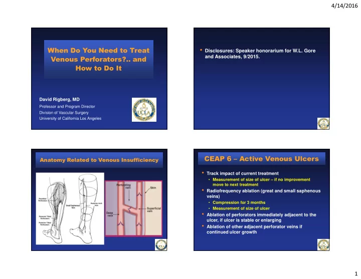
Anatomy Related to Venous Insufficiency

  • Track impact of current treatment
  • Measurement of size of ulcer – if no improvement

move to next treatment

  • Radiofrequency ablation (great and small saphenous

veins)

  • Compression for 3 months
  • Measurement of size of ulcer
  • Ablation of perforators immediately adjacent to the

ulcer, if ulcer is stable or enlarging

  • Ablation of other adjacent perforator veins if

continued ulcer growth

CEAP 6 – Active Venous Ulcers

slide-2
SLIDE 2

4/14/2016 2

  • Reversed Trendelenberg

position on an electronic tilt table

  • A GE Logiq-e portable duplex

scanner used for imaging

  • The incompetent vein adjacent

to the ulcer with both reflux and a diameter above 3 mm selected for Rx

Duplex Ultrasound Evaluation Duplex Ultrasound Evaluation Duplex Ultrasound Evaluation

  • RFS catheter placed at a 45°angle –

transverse and longitudinal transducer

  • Confirm stylet in vein at fascia
  • Inject local anesthetic to eliminate

pain

  • Trendelenberg position
  • Vein treated with RF energy for 1

minute in each quadrant; repeated above the fascia if possible

  • Confirmation of ablation due to

compression from local anesthetic

Duplex Ultrasound Evaluation

  • Leg dressed with compression

dressing

  • Confirmation of closure of the

vein at next clinic visit (48-72 hours postop)

  • Independent vascular lab

technician

  • Repeat procedure if incompletely

closed

slide-3
SLIDE 3

4/14/2016 3

Process of Perforator Closure

Learning curve of a Single Vascular Surgeon 0% 10% 20% 30% 40% 50% 60% 70% 80% 2007 2008 2009 2010 success rate

Perforator Closure Success Rate

0% 10% 20% 30% 40% 50% 60% 70% 80% 2007 2008 2009 2010

success rate

J Vasc Surg 2011;54(3):737-742

There Is A Learning Curve!

  • Ultrasound guided

foam sclerotherapy (UGFS) = 65%

  • Radiofrequency = 83%
  • Laser = 75%
  • SEPS = 87%

Mean Success Rate

*Studies in last 5 years Current State of the Treatment of Perforating Veins. JVS 2015.

Choosing the Right Modality for Perforator Ablation

0.00 2.00 4.00 6.00 8.00 10.00 12.00 14.00 16.00 0.00 0.10 0.20 0.30 0.40 0.50 0.60 Ulcer Size (cm^2) Time (Years) Ulcer growth = +2.66 cm2/mo Ulcer closure =

  • 6.08 cm2/mo

Green: Treatment with compression therapy alone Orange: Ongoing compression after vein ablation

Vein Ablation Procedure

Method of Tracking Ulcer Healing

slide-4
SLIDE 4

4/14/2016 4

Perforator Ablation for Venous Ulcers - RCT

Current State of the Treatment of Perforating Veins. JVS 2015. In-Press

  • Mean ulcer healing = 81%
  • Mean ulcer recurrence = 10%

J Vasc Surg 2012;55:458-464

Ablation Procedures Ulcer Locations

Impact of Perforator Ablation on Ulcer Healing Rates

Healed Ulcers N = 84 Healing Ulcers N = 26

Results – Change in Ulcer Healing Rate

Variable Pre-ablation healing rate (cm2/mo) ± ± ± ± SEM Post-ablation healing rate (cm2/mo) ± ± ± ± SEM P-value

Healed Ulcers ALL VEINS + 1.02 ± .11

  • 4.45 ± .13

<.001 Axial GSV + 1.18 ± .12

  • 6.44 ± .17

<.001 SSV + 1.06 ± .13

  • 4.82 ± .14

<.001 Perforators PTV + .89 ± .09

  • 2.92 ± .10

<.001 Unhealed Ulcers ALL VEINS + .96 ± .07

  • 4.60 ± .11

<.001 GSV + 1.09 ± .08

  • 6.26 ± .16

<.001 SSV + .99 ± .10

  • 4.57 ± .11

<.001 PTV + .80 ± .06

  • 2.79 ± .06

<.001

Ulcer Healing Rate by Specific Vein Ablation

slide-5
SLIDE 5

4/14/2016 5

  • Perforator ablation is difficult and takes practice
  • Venous ulcers can all be healed with an aggressive

approach to incompetent superficial axial veins, perforating veins, and occasionally correction on deep venous obstruction and/or reflux

  • The status of the ulcer is the key to determining if

there is “ambulatory venous hypertension”

  • If an ulcer in not healing with optimal compression or

heals and then recurs, there is a mechanical reason – we find it and treat it!

Conclusions