
SLIDE 1
12/1/17 1
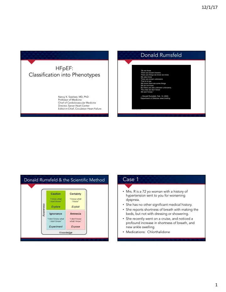
HFpEF: Classification into Phenotypes
Nancy K. Sweitzer, MD, PhD Professor of Medicine Chief of Cardiolovascular Medicine Director, Sarver Heart Center Editor-in-Chief, Circulation Heart Failure
Donald Rumsfeld
Donald Rumsfeld & the Scientific Method
Case 1
- Mrs. R is a 72 yo woman with a history of
hypertension sent to you for worsening dyspnea.
- She has no other significant medical history.
- She reports shortness of breath with making the
beds, but not with dressing or showering.
- She recently went on a cruise, and noticed a
profound increase in shortness of breath, and new ankle swelling
- Medications: Chlorthalidone