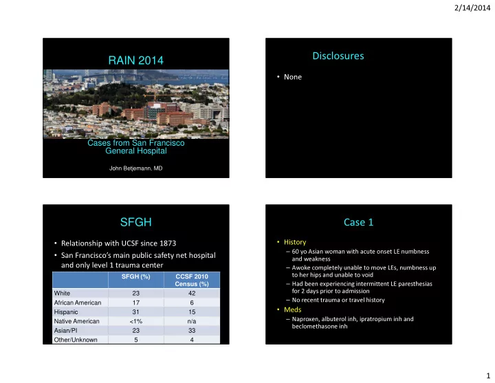
Disclosures RAIN 2014 None Cases from San Francisco General - - PowerPoint PPT Presentation

disclosures rain 2014
SMART_READER_LITE
LIVE PREVIEW

Disclosures RAIN 2014 None Cases from San Francisco General - - PowerPoint PPT Presentation

2/14/2014 Disclosures RAIN 2014 None Cases from San Francisco General Hospital John Betjemann, MD SFGH Case 1 History Relationship with UCSF since 1873 60 yo Asian woman with acute onset LE numbness San Franciscos main


slide-1
SLIDE 1

2/14/2014 1

RAIN 2014

Cases from San Francisco General Hospital

John Betjemann, MD

Disclosures

  • None

SFGH

  • Relationship with UCSF since 1873
  • San Francisco’s main public safety net hospital

and only level 1 trauma center

SFGH (%) CCSF 2010 Census (%) White 23 42 African American 17 6 Hispanic 31 15 Native American <1% n/a Asian/PI 23 33 Other/Unknown 5 4

Case 1

  • History

– 60 yo Asian woman with acute onset LE numbness and weakness – Awoke completely unable to move LEs, numbness up to her hips and unable to void – Had been experiencing intermittent LE paresthesias for 2 days prior to admission – No recent trauma or travel history

  • Meds

– Naproxen, albuterol inh, ipratropium inh and beclomethasone inh

slide-2
SLIDE 2

2/14/2014 2

Case 1

  • PMH

– Breast CA s/p lumpectomy and XRT in 2011 – Gastritis and asthma

  • Review of Systems

– 10 lb unintentional wt loss over last 1-2 weeks – Gastroenteritis 3 days prior to admission – Chronic cough with chest CT concerning for interstitial lung disease – Recent arthralgia in hands – Recent night sweats but no fevers – No issues with vision, speech or swallowing

Case 1: Exam

  • MS and CN exam unremarkable (including
  • phthalmologic exam)
  • Motor exam

– No pronator drift, symmetric fast finger taps – Flaccid paraparesis

  • No movement of LLE
  • Flicker of movement in proximal RLE and 4-/5 distally
  • Reflexes: 2+ in the UEs and absent in the LEs
  • Sensation: decreased to all modalities to nipple

line

  • Rectal tone was decreased

Case 1: Question 1

Which is the first diagnostic test you would like to perform?

C T b r a i n M R I b r a i n M R I C s p i n e M R I t

  • t

a l s p i n . . . L u m b a r p u n c t u r . . .

2% 0% 8% 83% 6%

  • 1. CT brain
  • 2. MRI brain
  • 3. MRI C spine
  • 4. MRI total spine
  • 5. Lumbar puncture

Case 1: Imaging

Sag T2 Sag T2 Sag T1 post con

slide-3
SLIDE 3

2/14/2014 3

Case 1: Question 2

Axial T2

What is your most likely diagnosis?

I n f e c t i

  • u

s ( v i . . . M u l t i p l e s c l e r . . . S a r c

  • i

d

  • s

i s N e u r

  • m

y e l i t i s . . . P a r a n e

  • p

l a s t i c . . . M i x e d c

  • n

n e c t i . . .

22% 0% 14% 41% 16% 6%

  • 1. Infectious (viral, TB etc.)

myelitis

  • 2. Multiple sclerosis
  • 3. Sarcoidosis
  • 4. Neuromyelitis optica
  • 5. Paraneoplastic myelitis
  • 6. Mixed connective tissue

disorder with autoimmune myelitis (i.e. Sjogren’s, lupus)

Case 1: Imaging and Labs

Axial FLAIR

  • CBC, Chem-7, LFTs normal
  • CSF
  • 161 RBC, 374 WBC (97%

neut), 194 protein, glucose 51 (120)

  • Serum ACE, dsDNA, ANA,

RF, RPR, B12, PPD and HIV all negative/normal

  • SSA/SSB, NMO, IgG index,
  • ligoclonal bands all

pending

Case 1: Question 3

Given the LP results, now what is your most likely diagnosis?

I n f e c t i

  • u

s ( v i . . . M u l t i p l e s c l e r . . . S a r c

  • i

d

  • s

i s N e u r

  • m

y e l i t i s . . . P a r a n e

  • p

l a s t i c . . . M i x e d c

  • n

n e c t i . . .

60% 0% 2% 20% 10% 8%

  • 1. Infectious (viral, TB etc.) myelitis
  • 2. Multiple sclerosis
  • 3. Sarcoidosis
  • 4. Neuromyelitis optica
  • 5. Paraneoplastic myelitis
  • 6. Mixed connective tissue disorder with

autoimmune myelitis (i.e. Sjogrens, Lupus)

Case 1: Question 4

Based on the current information, what is your next step?

S t a r t I V s

  • l

u m . . . S t a r t b r

  • a

d s p . . . I n i t i a t e I V I G P l a c e a l i n e f . . . A w a i t p e n d i n g . . .

35% 50% 2% 2% 10%

  • 1. Start IV solumedrol
  • 2. Start broad spectrum IV antibiotics

and acyclovir

  • 3. Initiate IVIG
  • 4. Place a line for plasma exchange
  • 5. Await pending labs prior to initiating

treatment

slide-4
SLIDE 4

2/14/2014 4

Case 1: Diagnosis

  • Initiated empiric solumedrol 1g IV x5 days
  • CSF:

– No oligoclonal bands – IgG index: 0.77 (0.23-0.64)

  • SSA (Ro) Ab: 156 (0-40 AU/mL)
  • SSB (La) Ab: 0 (0-40 AU/mL)
  • NMO ab: >160 U/mL (<4)

Case 1: Question 5

After 5 days of IV solumedrol the patient experienced minimal improvement. The preferred next step in management is?

P r e d n i s

  • n

e t a p . . . P r e d n i s

  • n

e t a p . . . P r e d n i s

  • n

e t a p . . . A n

  • t

h e r c

  • u

r s e . . . P r e d n i s

  • n

e t a p . . .

7% 23% 67% 2% 2%

  • 1. Prednisone taper and IVIG
  • 2. Prednisone taper and immunosuppression

(Rituximab)

  • 3. Prednisone taper and immunomodulators

(interferon)

  • 4. Another course of IV solumedrol
  • 5. Prednisone taper and plasma exchange

followed by immunosuppression (Rituximab)

Case 1: Follow up

  • s/p plasma exchange and 2 doses of Rituximab
  • MRI breast negative for tumor recurrence
  • 2.5 months out with no further attacks

– Ambulating with cane/walker – Has not regained bladder function

Case 1: Pearls

  • NMO

– Demographics: Asian and African Americans – Near complete LETM v. partial TM in MS – CSF: neutrophilic pleocytosis, eosinophils, relative absence of oligoclonal bands

  • NMO spectrum disorder

– Overlap with ANA and ENA (particularly SSA/SSB) – Demographics: female, median age at onset 56, more Caucasians – LETM more common than optic neuritis at presentation

slide-5
SLIDE 5

2/14/2014 5

Case 2

  • History

– 33 yo man presents with psychosis and suicidal ideation – 1 week prior to admission had intermittent LE paresthesias and numbness – 2-3 days prior to admission the LE symptoms worsened and he was unable to walk

  • Meds

– Noncompliant with risperidone and lorazepam

Case 2

  • PMH

– Suicide attempt in 9/12 – 1.5 years of paranoid delusions following the end

  • f a serious relationship

– History of polysubstance abuse (marijuana, ecstasy and methamphetamine) with rare etoh use

  • Review of Systems

– Recent weight loss but unclear how much – Otherwise unremarkable ROS

Case 2: Exam

  • MS: oriented, normal digit span, 0/3 recall
  • CN: unremarkable
  • Motor: normal bulk and tone. 5/5 throughout except

4+/5 in left triceps, interossei and hamstrings

  • Reflexes: 1+ in the UEs, 3 knees, 2+ ankles, toes down

going

  • Sens: decreased to vibration and proprioception

throughout (legs> arms). Pinprick and temperature intact

  • Coord: dysmetria in all 4 extremities with truncal ataxia
  • Gait: unable to stand without assistance

Case 2: Imaging

Sag T2 Axial FLAIR

slide-6
SLIDE 6

2/14/2014 6

Case 2: Question 1

What do you think will be most useful in guiding the differential diagnosis?

S e r u m l a b s L u m b a r p u n c t u r . . . M

  • r

e

  • f

a h i s t . . . A l l

  • f

t h e a b

  • .

. .

4% 90% 2% 4%

  • 1. Serum labs
  • 2. Lumbar puncture
  • 3. More of a history regarding

substance abuse

  • 4. All of the above

Case 2: SG

  • More history

– Patient admitted to using whippets twice daily for a year and recently had been attempting to overdose – “thousands of cartridges” covering his apartment

Case 2: Question 2

Which labs would you expect to be abnormal?

V i t B 1 2 H

  • m
  • c

y s t e i n e M e t h y l m a l

  • n

i c . . . P e r i p h e r a l s m e . . . A n s w e r s 1

  • 4

A n s w e r s 2

  • 4

4% 2% 24% 49% 4% 16%

  • 1. Vit B12
  • 2. Homocysteine
  • 3. Methylmalonic acid
  • 4. Peripheral smear
  • 5. Answers 1-4
  • 6. Answers 2-4

Case 2: Labs

  • CBC unremarkable

– Peripheral smear: hypersegmented neutrophils

  • Vit B12: 294 (236-888 pg/dL)

– was 618 on 9/19/12

  • Homocysteine: 197 (<10 umol/L)
  • Methylmalonic Acid: 3.85 (0-0.4)

– (1-9.99 consistent with mild B12 deficiency)

  • HIV, RPR, Chem-7 all unremarkable
slide-7
SLIDE 7

2/14/2014 7

Case 2: Question 3

How would you treat this patient?

S u p p

  • r

t i v e c a r . . . D a i l y

  • r

a l B 1 2 . . . D a i l y h i g h d

  • s

. . .

4% 91% 6%

  • 1. Supportive care
  • 2. Daily oral B12 supplementation
  • 3. Daily high dose IM B12

supplementation

Case 2: SG

  • No improvement in symptoms despite high

dose B12

– B12 level: 1108 pg/dL – Homocysteine 12 umol/L

Case 2: Repeat imaging

Sag T2 Axial T2

Case 2: Question 4

Would you perform an LP at this time

Y e s N

  • 30%

70%

  • 1. Yes
  • 2. No
slide-8
SLIDE 8

2/14/2014 8

Case 2: follow up

  • CSF

– 1 RBC, 0 WBC, gluc 60 mg/dL, prot 78 mg/dL – CSF gram stain and culture negative – Oligoclonal bands not detected – IgG index 0.52

  • Continued with high dose B12
  • supplementation. Patient has not returned

for follow up yet

Case 2: Pearls

  • N20 is an increasingly common drug of abuse

and is readily available

  • N2O causes the oxidation of Co+ to Co++,

thereby inactivating B12 in vivo

  • B12 is necessary for the synthesis of

methionine which is needed for myelin sheath protein

  • B12 levels may be normal but the patient is

functionally B12 deficient

Thank You

  • Questions?