Disclosure Diagnosis and Novel Management Strategies - - PDF document

disclosure
SMART_READER_LITE
LIVE PREVIEW

Disclosure Diagnosis and Novel Management Strategies - - PDF document

Pulmonary Arterial Hypertension: Disclosure Diagnosis and Novel Management Strategies Grants/Research Support: 2016 Lung Biotechnology, Pfizer, Reata Teresa De Marco, MD, FACC Consultant Professor of Medicine & Surgery


slide-1
SLIDE 1

1

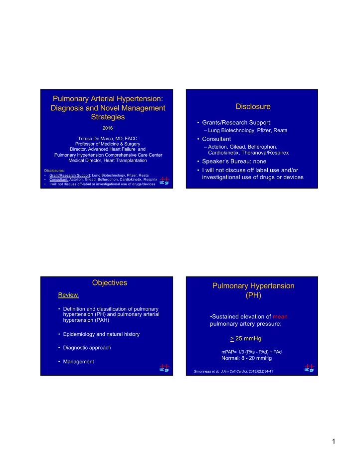
Pulmonary Arterial Hypertension: Diagnosis and Novel Management Strategies

2016 Teresa De Marco, MD, FACC Professor of Medicine & Surgery Director, Advanced Heart Failure and Pulmonary Hypertension Comprehensive Care Center Medical Director, Heart Transplantation

Disclosures:

  • Grant/Research Support: Lung Biotechnology, Pfizer, Reata
  • Consultant: Actelion, Gilead, Bellerophon, Cardiokinetix, Respirix
  • I will not discuss off-label or investigational use of drugs/devices

UC UC SF SF

Disclosure

  • Grants/Research Support:

– Lung Biotechnology, Pfizer, Reata

  • Consultant

– Actelion, Gilead, Bellerophon, Cardiokinetix, Theranova/Respirex

  • Speaker’s Bureau: none
  • I will not discuss off label use and/or

investigational use of drugs or devices

Objectives

Review:

  • Definition and classification of pulmonary

hypertension (PH) and pulmonary arterial hypertension (PAH)

  • Epidemiology and natural history
  • Diagnostic approach
  • Management

UC UC SF SF

Pulmonary Hypertension (PH)

  • Sustained elevation of mean

pulmonary artery pressure: > 25 mmHg

UC UC SF SF Simonneau et al, J Am Coll Cardiol. 2013;62:D34-41

mPAP= 1/3 (PAs - PAd) + PAd

Normal: 8 - 20 mmHg

slide-2
SLIDE 2

2

5th WSPH Updated Classification of Pulmonary Hypertension, Nice 2013

GROUP 1 – Pulmonary Arterial Hypertension

1.1 Idiopathic PAH 1.2 Heritable PAH 1.2.1 BMPR2 1.2.2 ALK-1, ENG, SMAD9, CAAV1, KCNK3 1.2.3 Unknown 1.3 Drug- and Toxin-Induced 1.4 Associated with: 1.4.1 Connective Tissue Disease 1.4.2 Human Immunodeficiency Virus (HIV) Infection 1.4.3 Portal Hypertension 1.4.4 Congenital Heart Disease 1.4.5 Schistosomiasis 1’ Pulmonary Veno-occlusive Disease and/or Pulmonary Capillary Hemangiomatosis 1” Persistent Pulmonary Hypertension of the Newborn (PPHN)

GROUP 2 – PH Due to Left Heart Disease

2.1 Left Ventricular Systolic Dysfunction 2.2 Left Ventricular Diastolic Dysfunction 2.3 Valvular Disease 2.4 Congenital/Acquired Left Heart Inflow/Outflow Tract Obstruction and Congenital Cardiomyopathies

GROUP 3 –PH Due to Lung Disease and/or Hypoxia

3.1 Chronic Obstructive Pulmonary Disease 3.2 Interstitial Lung Disease 3.3 Other Pulmonary Diseases W ith Mixed Restrictive and Obstructive Pattern 3.4 Sleep-disordered Breathing 3.5 Alveolar Hypoventilation Disorders 3.6 Chronic Exposure to High Altitude 3.7 Developmental Lung Diseases

GROUP 4 – Chronic Thromboembolic PH (CTEPH) GROUP 5 – PH With Unclear Multifactorial Mechanisms

5.1 Hematologic Disorders: Chronic Hemolytic Anemia, Myeloproliferative Disorders, Splenectomy 5.2 Systemic Disorders: Sarcoidosis, Pulmonary Histiocytosis, Lymphangioleiomyomatosis 5.3 Metabolic Disorders: Glycogen Storage Disease, Gaucher Disease, Thyroid Disorders 5.4 Others: Tumoral Obstruction, Fibrosing Mediastinitis, Chronic Renal Failure, Segmental PH

Simmoneau G, et al. JACC. 2013; 62:D34-41. BMPR2 = bone morphogenetic protein receptor type 2; CAV1 =caveolin 1; ENG = endoglin; KCNK3 = gene encoding K2P3.1 (K+ channel)

Strange G et al. Heart 2012;98:1806-1811.

  • Single center study from Australia
  • 6,994 screened à 936 pts (9.1%) with PH on ECHO

(defined as ePASP >40 mmHg)

Etiology of PH on Echocardiogram 5th WSPH Clinical Classification of PAH

(WHO Group 1)

Group 1―Pulmonary Arterial Hypertension (PAH)

Idiopathic PAH Heritable BMPR2 ALK-1, endoglin, SMAD9,CAAV1,KCNK3 Unknown Drug and toxin-induced PAH associated with: Connective tissue disease HIV infection Portal hypertension Congenital heart disease Schistosomiasis 1’ – Pulmonary veno-occlusive disease or pulmonary capillary hemangiomatosis 1’’ – Persistent PH of the newborn

Simonneau et al, J Am Coll Cardiol. 2013;62:D34-41.

Group 1: Pulmonary Arterial Hypertension (PAH)

  • Characterized by progressive and sustained elevation of

pulmonary artery pressure and vascular resistance: – PA mean > 25 mmHg (nl 8-20 mmHg) – PAWP/LVEDP < 15 mmHg (nl 4-12 mmHg) – PVR > 3 W units (240 dyn/sec/cm-5)

  • Subset of PH (15 cases/ million)
  • US prevalence 50-100,000
  • 15 – 25,000 dx & rx
  • Vasoconstriction, remodeling, thrombosis in situ
  • Progressive cardiopulmonary deterioration
  • Leads to RH failure and death (67% 5-yr survival)

Hoeper MM, et al. J Am Coll Cardiol. 2013;62:D42-50. Humbert M et al. Am J Respir Crit Care Med 2006;173:1023-30 Thenappan T, et al. Eur Respir J. 2010;35:1079-1087.

slide-3
SLIDE 3

3

NORMAL REVERSIBLE DISEASE IRREVERSIBLE DISEASE

Pathogenesis of Pulmonary Arterial Hypertension

Gaine S, J Am Med Assoc 2000;284:3160-68

Survival of PAH Patients in Current Era: Comparison with Historical Controls

Thenappan T, et al. Eur Respir J. 2010;35:1079-1087.

67 65 32 100 91 76 43 10 20 30 40 50 60 70 80 90 100 Baseline 1 year 3 year 5 year

Percentage (%) Survival

Observed Predicted (NIH)

N = 276, IPAH and HPAH patients diagnosed from 1982-2006; matched for disease variables at baseline with historical controls Low Risk Determinants of Risk High Risk

No Clinical evidence of RV failure Yes Gradual Disease progression Rapid II, III WHO functional class IV Longer (> 400 meters) 6-MWD Shorter (< 300 meters) Peak VO2 > 10.4 mL/kg/min Cardiopulmonary exercise testing Peak VO2 < 10.4 mL/kg/min Minimally elevated and stable BNP/NT-proBNP Significantly elevated PaCO2 > 34 mm Hg Blood gasses PaCO2 < 32 mm Hg Minimal RV dysfunction ECHO findings Pericardial effusion, RV dysfunction, RA enlargement RAP < 10 mm Hg; CI > 2.5 L/min/m2 Hemodynamics RAP > 20 mm Hg; CI < 2 L/min/m2

McLaughlin, et al. J Am Coll Cardiol 2009;53:1573 D’Alonzo, et al. Ann Int Med 1991;115:343 Raymond, et al. J Am Coll Cardiol 2002;39:1214

PAH Determinants of Patient Risk

and Prognosis

ACC/AHA Expert Consensus

Provencher, et al. E Heart J 2006: 27:589 Nagaya, et al. Circ 2000;102:865 Blyth, et al. Eur Respir J. 2007;29:737

Hemodynamic Changes Correlate with Disease Progression

CO=cardiac output; PAP=pulmonary arterial pressure; PVR=pulmonary vascular resistance; RAP=right atrial pressure.

Time PAP PVR CO Presymptomatic/ Compensated Symptomatic/ Decompensating Symptom Threshold

Right Heart

Declining/ Decompensated RAP

slide-4
SLIDE 4

4

Evaluation

UC UC SF SF

REVEAL Database: Most Frequent Symptoms at Diagnosis

Elliott EG, et al. Chest. 2007;132(4 suppl):631S.

Dyspnea at rest Cough Dizzy/lightheaded Presyncope/syncope Edema Chest pain/discomfort Other Fatigue Dyspnea on exertion

84.0% 26.0% 24.0% 23% 21.0% 23.0% 16.0% 13.0% 11% 83% 29% 27% 20% 20% 20% 14% 13% 11%

0 25 50 75 100 Incidence (%) IPAH APAH

N=1479.

PAH Diagnostic Guidelines: Decision Analysis

McGoon M, et al. Chest. 2004;126:14S-34S.

Unexplained Symptoms of Dyspnea on Exertion, Syncope/Near Syncope, Fatigue Clinical History, Examination, ECG, Chest X-Ray

Signs on Physical Examination

  • Loud pulmonic valve closure (P2) (93%)
  • TR murmur (40%)
  • PR murmur (13%)
  • Right-sided fourth heart sound
  • Right ventricular lift
  • Jugular venous distention
  • RV third heart sound (23%)
  • Peripheral edema (32%)
  • Ascites
  • Low BP, low PP, cool extremities (low CO,

peripheral vasoconstriction, hypoperfusion)

  • Stigmata of associated causes of PAH

Signs of RHF

McLaughlin VV et al. J ACC. 2009;53:1573-1619 McGoon M, et al. Chest. 2004;126:14S−34S. Rich S, et al. Ann Intern Med. 1987;107:216-223.

UC UC SF SF

slide-5
SLIDE 5

5

Electrocardiogram

Image courtesy of Vallerie McLaughlin, MD

  • Insufficiently sensitive as screening tool for PH
  • Prognosis:­p-wave in II, qR V1, RVHè­risk of death
  • RAD, RAE, RBBB,RVH

Bossone E, et al. Chest 2002;121:513 McGoon M, et al. Chest 2004;126:14S-34S McLaughlin VV et al. JACC 2009;53:1573-1619.

Prominent Central Pulmonary Artery Peripheral Hypovascularity Right Descending Pulmonary Artery RV Enlargement §• Cardiac enlargement §• Prominent proximal PA s §• “Pruning” of distal PA s § • No evidence of pulmonary edema § • Lungs appear normal

Chest Radiograph in PAH

PAH Diagnostic Guidelines: Decision Analysis

McGoon M, et al. Chest. 2004;126:14S-34S.

Clinical History, Examination, Chest X-Ray, ECG Is There a Reason to Suspect PH? Yes No Echocardiography Work-Up for Other Conditions

LV RV

Apical Four Chamber Normal PAH

LV RV RA LA

Parasternal Short Axis

slide-6
SLIDE 6

6

Signs of PAH with Echo/Doppler

  • Increased sPAP or TR jet
  • Right atrial and ventricular

hypertrophy/enlargement

  • Flattening of intraventricular

septum

  • Tricuspid regurgitation
  • Small LV dimension

LV RV LA RA IVS

ePAsP = 4V2 + eRAP

Traditional ECHO does not accurately measure:

  • Mean PA pressure
  • PAWP
  • Cardiac output (blood flow)
  • Cannot calculate PVR
  • Other limitations-15% no TR jet,

not all congenital lesions obvious, small errors in TRV tracing can alter results

McGoon M, et al. Chest. 2004;126:14S−34S

  • Contraction of RV is mainly longitudinal; tricuspid annulus displaced

toward apex during systole

  • Imaging through lateral wall with M-mode to measure this motion known

as tricuspid annular plane systolic excursion (TAPSE)

  • Less displacement as RV becomes more dysfunctional
  • Baseline TAPSE <1.8 cm has negative prognostic implications

Forfia PR et al. Am J Respir Crit Care Med. 2006;174:1034-1041.

RV function: Tricuspid Annular Plane

Systolic Excursion

Normal RVF: 2.3 – 2.7 cm Mild RVD: 1.9 – 2.2 cm Moderate RVD: 1.5 - 1.8 cm Severe RVD: <1.5 cm

PAH Diagnostic Guidelines

McGoon M, et al. Chest. 2004;126:14S-34S.

Echocardiography Indicates PH

Evaluate for Associated Causes V/Q scan PFTs Arterial Saturation

HIV Infection, Scleroderma, SLE, Other CTD, Liver Disease, CHD, Drug- Associated Parenchymal Lung Disease, Hypoxemia,

  • r Sleep Disorder

Suspected Chronic PE

slide-7
SLIDE 7

7

Ventilation Perfusion Lung Scan

Idiopathic PAH Chronic PE

Ventilation Perfusion Perfusion Ventilation

Ø 3 – 4% of acute PE do not entirely resolve Ø 50% of those with CTEPH do not have hx of acute PE Ø V/Q scan is sensitive, should be performed to exclude CTEPH even when another explanation for PH is present Ø CTEPH: >1 segmental-sized or larger mismatched perfusion defects Ø Normal or very low probability V/Q scan excludes CTEPH

Bands Absent branches Pouch

CTEPH: A “Curable” Form of PH Not to Be Missed

PAH Diagnostic Guidelines

McGoon M, et al. Chest. 2004;126:14S-34S.

Echocardiography Indicates PH

Evaluate for Associated Causes V/Q scan PFTs Arterial Saturation

HIV Infection, Scleroderma, SLE, Other CTD, Liver Disease, CHD, Drug- Associated Parenchymal Lung Disease, Hypoxemia,

  • r Sleep Disorder

Suspected Chronic PE

Pulmonary Function Tests, Arterial Blood Gases, and Oxygen Saturation

  • Findings suggestive
  • f PAH

– ê DLCO 40% - 80% of expected – Mild to moderate ê

  • f

lung volumes – Peripheral airway

  • bstruction

– Arterial O2 tension normal

  • r slightly ê at rest

– Arterial CO2 is ê – SpO2 preserved at rest, may be ê with exercise/ambulation

  • Findings suggestive of

alternate PH diagnoses – Hypoxic PH due to COPD

  • Irreversible airway
  • bstruction + increased

residual volumes

  • reduced DLCO + normal or

increased CO2 tension

– Interstitial lung disease

  • Decrease in lung volume +
  • Decreased DLCO

Galie N, et al. Eur Heart J. 2009;30(20):2493-2537.

slide-8
SLIDE 8

8

PAH Diagnostic Guidelines: Confirmation of PAH

Adapted from McGoon M, et al. Chest. 2004;126:14S-34S.

Echocardiography Indicates PH Right Heart Catheterization

  • Establish diagnosis
  • Ascertain etiology
  • Establish severity & prognosis
  • Verify presence and severity of shunts
  • Evaluate vasoreactivity
  • Guide treatment

VC RA RV PA PV PC LA LV Ao

Isolated post-capillary (Passive PH )

PH Hemodynamic Profiles:

Where is the lesion? (mean PAP > 25 mmHg) Combined post- pre-capillary PH (Mixed PH) Pre-capillary PH High flow PH

nl range or éPVR (CHD, AV fistula, thyrotoxicosis, chronic anemia) PAWP: Normal 4-12 mmHg TPG= mPAP - PAWP Normal < 10 mmHg DPG= dPAP – PAWP Normal < 5 mmHg PVR= mPAP - PAWP/CO Normal < 1 Wu

Vachiery JL et al, J Am Coll Cardiol 2013;62: D100-8 Fang J et al, J Heart Lung Transplant 2012;31:913–33 Galie N et al, Eur Respir J 2009;34(6):1219-63

Algorithm for Assessment of Vasoreactivity in Patients with PAH

Right Heart Catheterization With Acute Vasoreactivity Testing (iNO, epoprostenol, adenosine) Non - responder Consider: Oral ERAs/PDEI-5/sGCS/IPA Inhaled Iloprost SQ/IV/inhaled/PO Treprostinil IV Epoprostenol Responder (<15%) Consider: Hemodynamically- Monitored Trial of Calcium Channel Blocker

mPA ¯10 mmHg ¯mPA < 40 mmHg No D CO

(<10% respond long-term)

PAH Diagnostic Workup

McGoon M, et al. Chest. 2004;126:14S-34S.

Right Heart Catheterization Confirms PAH

6-minute walk, Borg score NYHA/WHO functional class

Establish Baseline, Prognosis, and Document Progression/Response to Treatment With Serial Re-assessment

slide-9
SLIDE 9

9

Management

UC UC SF SF

Goals of Management of PAH Prevent right heart failure

  • Alleviate symptoms
  • Improve exercise capacity
  • Improve functional class
  • Improve hemodynamics
  • Prevent clinical worsening
  • Reduce morbidity, mortality

Overview: 2013 WSPH Treatment Algorithm

Adapted from Galié N et al JACC. 2013;62 (25,

  • Suppl. D):D60-D72.

General Measures and Supportive Therapy Expert Referral (PH Center) Acute Vasoreactivity Testing Oral Anticoagulants IPAH, heritable PAH anorexigen-induced PAH APAH Diuretics Oxygen Digoxin (controversial)

Non-Vasoreactive

WHO-FC I-III CCB Sustained Response (WHO FC I-II) Continue CCB YES

Initial Therapy With PAH Approved Drugs

Depending on WHO-FC Status and risk profile

Vasoreactive

NO

Supervised Exercise Training Psychosocial Support Avoidance of Strenuous Activity Pregnancy Avoidance Immunizations (influenza)

Therapeutic Targets for PAH

Modified from Humbert M, Sitbon O, Simonneau G. N Engl J Med 2004;351:1425-36 Phosphodiesterase type 5 inhibitor Exogenous nitric oxide sGC stimul. Endothelin receptor antagonists Prostacyclin derivatives IP rec. agonist Endothelin receptor A

Endothelin 1 Nitric oxide Prostacyclin (prostaglandin I2)

Endothelin receptor B

Vasodilation and antiproliferation Vasodilation and antiproliferation Vasoconstriction and proliferation cGMP cAMP

Pre-proendothelin à Proendothelin L-arginine à L-citrulline Arachidonic acid à Prostaglandin I2

+ +

Phosphodiesterase type 5 Smooth muscle cells Endothelin cells Vessel lumen

Nitric oxide pathway Endothelin pathway Prostacyclin pathway

slide-10
SLIDE 10

10

FDA -Approved Specific Therapies for PAH

Class of Agent Name Route of Administration Oral Inhaled IV/SC Endothelin Receptor Antagonist Ambrisentan ✔ Bosentan ✔ Macitentan ✔ Phosphodiesterase Type 5 Inhibitor Sildenafil ✔ Tadalafil ✔ Soluble Guanylate Cyclase Stimulator Riociguat ✔ Prostacyclin Receptor Agonist Selexipag ✔ Prostacyclin analog Epoprostenol IV Iloprost ✔ Treprostinil ✔ ✔ IV and SC

US Food and Drug Administration. Approved product list as of December 22, 2015.

Intravenous epoprostenol Subcutaneous treprostinil Inhalational Iloprost Inhalational Treprostinil

2015 ESC/ERS Guidelines: Treatment Algorithm for PAH-specific Therapy

Galie N, et al. Eur Respir J. 2015;46:903-975.

Low or intermediate risk (WHO FC II-III) High risk (WHO FC IV)

Inadequate clinical response Consider referral for lung transplantation Inadequate clinical response

Patient already

  • n treatment

Initial monotherapy* Initial oral combination

Initial combination including IV prostacyclin

Double or triple sequential combination Consider listing for lung transplantation

*Initial combination with ambrisentan plus tadalafil has proven to be superior to monotherapy in delaying clinical failure.

5th WSPH: Prognostic Variables Used in Clinical Practice To Set Treatment Goals

Variable Recommended Goal

NYHA Functional class I or II Echocardiography/CMR Normal/near normal RV size and function Hemodynamics Normalization of RV function

  • RAP < 8 mm Hg and
  • CI > 2.5 to 3.0 L/min/m2

6 Minute walk distance Cardiopulmonary exercise testing >380-440 m (or more in younger pts) Peak VO2 >15 mL/min/kg and EqCO2 <45 L/min/min B-type natriuretic peptide Normal range

McLaughlin VV, et al. J Am Coll Cardiol. 2013;62:D73-81.

slide-11
SLIDE 11

11

Inadequate Clinical Response to Initial PAH Therapy

Failure to show improvement or deterioration with monotherapy

Consider eligibility for lung transplant

Inadequate Clinical Response on Maximal Therapy? Lung transplant (I-C) BAS (Iia- C)

Adapted from Galié N et al JACC. 2013;62 (25):D60-D72.

GRIPHON: Selexipag for PAH Primary Endpoint: Time to First Event

Placebo 582 433 347 220 149 88 28 Selexipag 574 455 361 246 171 101 40

  • No. at Risk:

Months

Patients without an event (KM)%

Selexipag vs placebo: RR 40% HR = 0.60; 99% CI, 0.46-0.78 P<0.0001 Selexipag Placebo

Sitbon O, et al. N Engl J Med. 2015:373:2522-2533.

At baseline: 20% PAH therapy naive 47% on monotherapy (ERA or PDE-5i) 33% on combination therapy (ERA & PDE-5i)

New Paradigm- AMBITION: Ambrisentan-Tadalafil Up-front Combination Therapy

Primary Endpoint: Time to First Clinical Failure Event Primary Analysis Set Event-Free (%) Time (weeks)

HR: 0.502 95% CI(0.348, 0.724) p=0.0002

0 24 48 72 96 120 144 168 192 1 year 88.9% 1 year 75.5% Combination therapy Pooled monotherapy 2 year 79.7% 2 year 63.2% 3 year 56.1% 3 year 67.6% Combination: 253 229 186 145 106 71 36 4 Pooled monotherapy: 247 209 155 108 77 49 25 5

Number at risk: Galie N, et al. N Engl J Med. 2015;373:834-844.

PAH: Therapy

Transplantation - lung / heart-lung

  • Indicated for patients who continue to deteriorate

with poor QOL despite aggressive therapy

  • 1 year survival – 70-90%
  • 5 year survival – 50-60%
  • Mortality on waiting list remains high

(20% per yr despite LAS)

  • Aggressive pre-transplant management can

ê mortality

UC UC SF SF

Chen H … De Marco et al. Am J Resp Crit Care Med 2009;180:468

slide-12
SLIDE 12

12

PAH: Summary

  • PAH is a progressive disease associated with

significant morbidity and mortality

  • Echocardiography and right heart catheterization

are the primary diagnostic modalities

  • Strides

made thus far in pathogenesis and pathobiology have lead to more effective therapies

  • Current therapies have significant limitations and

are costly

  • New

therapeutic agents and strategies are available and emerging