DISCLOSURE: Joseph Sowka, OD is/ has been a Consultant/ Speaker - - PDF document

disclosure
SMART_READER_LITE
LIVE PREVIEW

DISCLOSURE: Joseph Sowka, OD is/ has been a Consultant/ Speaker - - PDF document

2/6/2019 DISCLOSURE: Joseph Sowka, OD is/ has been a Consultant/ Speaker Bureau/ Advisory Board member for Novartis, Allergan, Glaukos, and B&L. Dr. Sowka has no direct financial interest in any of the diseases, products or


slide-1
SLIDE 1

2/6/2019 1

NEURO-OPHTHALMIC UPDATE

Joseph Sowka, OD Greg Caldwell, OD

Joseph Sowka, OD is/ has been a Consultant/ Speaker Bureau/ Advisory Board member for Novartis, Allergan, Glaukos, and B&L. Dr. Sowka has no direct financial interest in any of the diseases, products

  • r

instrumentation mentioned in this presentation. He is a co-owner

  • f

Optometric Education Consultants

The ideas, concepts, conclusions and perspectives presented herein reflect the opinions of the speaker; he has not been paid, coerced, extorted or otherwise influenced by any third party individual or entity to present information that conflicts with his professional viewpoints.

DISCLOSURE:

DISCLOSURES- GREG CALDWELL, OD, FAAO

Will mention many products, instruments and companies during our discussion

  • I don’t have any financial interest in any of these products, instruments or

companies Pennsylvania Optometric Association –President 2010

  • POA Board of Directors 2006-2011

American Optometric Association, Trustee 2013-2016

  • Thank you to the members and those who join

I never used or will use my volunteer positions to further my lecturing career Lectured for: Shire, BioTissue, Optovue Advisory Board: Allergan Envolve: PA Medical Director, Credential Committee

He is a co-owner of Optometric Education Consultants
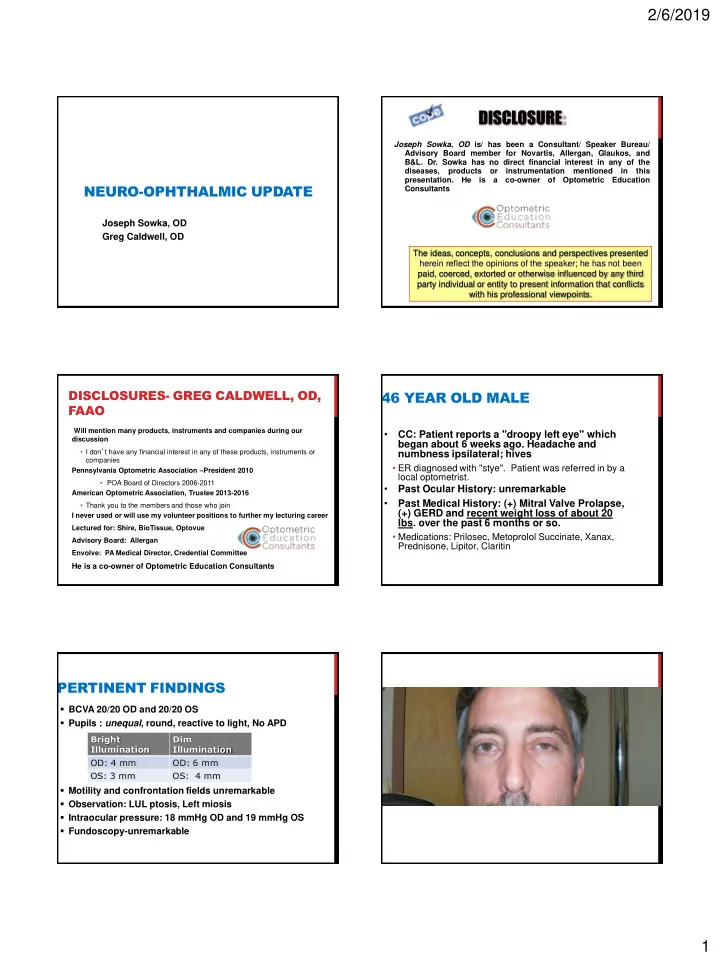
46 YEAR OLD MALE

  • CC: Patient reports a "droopy left eye" which

began about 6 weeks ago. Headache and numbness ipsilateral; hives

  • ER diagnosed with "stye". Patient was referred in by a

local optometrist.

  • Past Ocular History: unremarkable
  • Past Medical History: (+) Mitral Valve Prolapse,

(+) GERD and recent weight loss of about 20

  • lbs. over the past 6 months or so.
  • Medications: Prilosec, Metoprolol Succinate, Xanax,

Prednisone, Lipitor, Claritin

PERTINENT FINDINGS

  • BCVA 20/20 OD and 20/20 OS
  • Pupils : unequal, round, reactive to light, No APD
  • Motility and confrontation fields unremarkable
  • Observation: LUL ptosis, Left miosis
  • Intraocular pressure: 18 mmHg OD and 19 mmHg OS
  • Fundoscopy-unremarkable

Bright Illumination Dim Illumination OD: 4 mm OD: 6 mm OS: 3 mm OS: 4 mm

slide-2
SLIDE 2

2/6/2019 2

So, what do you think and what do you want to do now?

POST-IOPIDINE

Pre-Iopidine Post-Iopidine

HORNER’S SYNDROME

  • Etiology unclear based upon exam
  • Headache, neuralgia and ‘hives’
  • Not consistent with cluster migraine
  • Dx of exclusion, not convenience
  • Hives- not consistent with HZO
  • Unexplained weight loss concerning-

relationship unclear

  • Recommend medical eval by PCP
  • Additional testing dictated by PCP results

DISCUSSION

What is Horner’s Syndrome?

  • a triad of clinical signs arising from disruption
  • f sympathetic innervation to the eye and

ipsilateral face that causes miosis, upper lid ptosis, mild elevation of the lower lid, and anhydrosis of the facial skin.

slide-3
SLIDE 3

2/6/2019 3

PHARMACOLOGICAL TESTING

  • Cocaine
  • Horner’s pupil doesn’t dilate, normal pupil does
  • Hydroxyamphetamine (Paredrine)
  • Differentiates post- from pre-ganglionic
  • Not available and doesn’t matter because bad stuff

happens everywhere

  • Apraclonidine 0.5% (Iopidine)
  • Denervation suprasensitivity
  • 36-72 hours from onset
  • Horner’s pupil dilates, normal doesn’t
  • Reversal more classic and diagnostic that cocaine

HORNER’S SYNDROME: ETIOLOGIES

First-order neuron disorder: Stroke (e.g., vertebrobasilar artery insufficiency or infarct); tumor; multiple sclerosis (MS), and, rarely, severe osteoarthritis of the neck with bony spurs. Second-order neuron disorder: Tumor (e.g., lung carcinoma, metastasis, thyroid adenoma, neurofibroma). Patients with pain in the arm or scapular region should be suspected of having a Pancoast tumor. In children, consider neuroblastoma, lymphoma, or metastasis.

HORNER’S SYNDROME: ETIOLOGIES

  • Third-order neuron disorder: Headache syndrome

(e.g., cluster, migraine, Raeder paratrigeminal syndrome), internal carotid dissection, herpes zoster virus, otitis media, Tolosa–Hunt syndrome, neck trauma/tumor/inflammation, prolactinoma.

  • Congenital Horner syndrome: Trauma (e.g., during

delivery).

  • Facebook tomography
  • Other rare causes: Cervical paraganglioma, ectopic

cervical thymus

MANAGEMENT

  • Localizable- targeted workup
  • Neck and facial pain- carotid dissection
  • Facial paraesthesia- middle cranial fossa disease
  • Necessary Work Up (non-localizable):
  • MRI of brain, orbits and chiasm with and without contrast,

attention to middle cranial fossa.

  • MRA of head and neck-rule out carotid dissection
  • MRI of neck and cervical spine, include lung apex and brachial

plexus

  • Horner’s syndrome patient needs to be imaged from chest to head- 3

scans

  • Horner’s protocol
  • All imaging in patient unremarkable

CAROTID DISSECTION

 A 3rd-order Horner’s and ipsilateral head, eye, or neck pain of acute onset should be considered diagnostic of internal carotid dissection unless proven

  • therwise.

CAROTID DISSECTION

  • Carotid artery dissection presents with the

sudden or gradual onset of ipsilateral neck or hemicranial pain, including eye or face pain

  • Often associated with other neurologic findings

including an ipsilateral Horner’s syndrome, TIA, stroke, anterior ischemic optic neuropathy, subarachnoid hemorrhage, or lower cranial nerve palsies

  • 52% with ocular or hemispheric stroke with 6 days
  • 67% within first week; 89% within 2 weeks; none after 31 days
  • Horner’s from suspected carotid dissection

should go to ER

slide-4
SLIDE 4

2/6/2019 4

HORNER SYNDROME ALGORITHM

  • 1. Confirm it is Horner syndrome
  • Apraclonidine; dilation lag
  • 2. Determine if accidental or surgical trauma

as cause

  • 3. Urgent imaging
  • CT/CTA; MRI/MRA head and neck if present< 2 weeks
  • 4. Image lung apex

RULE Diagnosing Horner’s syndrome is insufficient. You must try to ascertain a cause and never assume that it is benign. CASE: 59 BF

  • Long time patient presents for her glaucoma

f/u. She reports drooping in the right eye and smaller pupil for about 1 month. Symptoms were noticed at/ about time of dx of lung cancer and subsequent surgery.

  • `She also reports scapular pain and weakness in the

right hand.

  • Past Medical History: (+) Lung Cancer, (+)

Pancreatitis, (+) HTN and (+) Acid Reflux

  • Social History: Smokes 1 pack per day for 45

years, Drinks a 6 pack of beer daily

CASE: PERTINENT FINDINGS CONTINUED…

  • Pharmacological testing not done
  • New onset of ptosis and miosis with

dx lung cancer and h/o recent lung surgery

  • Dx=Pancoast Syndrome

PANCOAST TUMOR

A Pancoast tumor is a lung cancer arising in the apex of the lung that involves structures of the apical chest wall. Treatment

  • Chemotherapy
  • Radiation Therapy
  • Surgery: lobectomy vs. wedge resection

Prognosis: 5 year survival rate is around 30%

  • Not an emergency
slide-5
SLIDE 5

2/6/2019 5

ODE TO HORNER’S SYNDROME

When the lid is low and the pupil small, Check to see the sweat don’t fall. Cocaine is no longer universal, Iopidine will cause reversal. You have to scan head to chest, And remember that MRA is best. Pain in association, will surely cause commotion. Send to the ER without correction, Remember, it might be carotid dissection.

Joseph Sowka, OD

80 YEAR OLD MAN

  • Reports a sudden loss of vision OD
  • Vision is count fingers at 2 feet OD and 20/25

OS

  • APD OD grade 4
  • Fundus photos OU

PHOTOS OU

CRAO TREATMENT/WORK- UP/FOLLOW-UP?

  • Anterior chamber paracentesis (less than 24

hours)

  • STAT blood work
  • 2-10% of all CRAOs are caused by thrombosis from

Giant Cell Arteritis (GCA)

  • Sed-rate
  • C-reactive protein
  • Qualitative or quantitative?
  • CBC with diff
  • Monitor for neovascularization, every 3-6

weeks❌ 🆙

CRAO, BRAO, TIA (AMAUROSIS

FUGAX)

  • Acute Stroke Ready Hospital
  • Certification recognizes hospitals that meet standards to support better outcomes for stroke care as part of a stroke system of

care

  • Developed in collaboration with the Joint Commission (TJC), eligibility standards include:
  • Dedicated stroke-focused program
  • Staffing by qualified medical professionals trained in stroke care
  • Relationship with local emergency management systems (EMS) that encourages training in field assessment tools

and communication with the hospital prior to bringing a patient with a stroke to the emergency department

  • Access to stroke expertise 24 hours a day, 7 days a week (in person or via telemedicine) and transfer agreements with facilities

that provide primary or comprehensive stroke services.

  • 24/7 ability to perform rapid diagnostic imaging and laboratory testing to facilitate the administration for IV thrombolytics in

eligible patients

  • Streamlined flow of patient information while protecting patient rights, security and privacy
  • Use of data to assess and continually improve quality of care for stroke patients
  • Warn hospital is suspicion for GCA
  • 20% of stroke or heart attack within 3 years
  • However of those who experienced CVA or MI
  • 80% were within 24-48 hours; those remaining
  • 50% occurred in 2 weeks
  • Majority within the next 90 days
  • Not PCP, not retinologist, just the Acute Stroke Ready Hospital!

35 YEAR OLD MAN

  • Wants another opinion due to “hemorrhage
  • n my right eye”
  • Happened 3 days ago after vomiting
  • Claims food poisoning from chicken Caesar salad
  • Still feels a little nauseated
  • Saw ophthalmologist 3 days ago, told he had

a bruise on his eye and it should go away in 1-2 weeks

slide-6
SLIDE 6

2/6/2019 6

35 YEAR OLD MAN

BVA 20/100 OD, 20/70 OS

  • Hx of amblyopia OD
  • Current Rx OD +5.50 OS +4.50

Any concerns? Patient noticed blurry vision OS

  • Started 2 weeks ago
  • Did not mention because he is more

concerned about the blood on his right eye

Headaches for 2 weeks, decrease if patient stands up ROS: unremarkable Decide to dilate OU

RETINAL FINDINGS DISCUSSION DIFFERENTIAL DIAGNOSIS

  • Hypertensive retinopathy
  • Blood dyscrasia
  • Terson’s syndrome
  • Valsalva retinopathy
  • Purtscher’s retinopathy
  • Shaken baby syndrome

TERSON’S SYNDROME

  • Terson’s syndrome originally was defined by

the occurrence of vitreous hemorrhage in association with subarachnoid hemorrhage.

  • Terson’s syndrome now encompasses any

intraocular hemorrhage associated with intracranial hemorrhage and elevated intracranial pressures.

  • Intraocular hemorrhage includes the

development of subretinal, retinal, subhyaloidal, or vitreal blood.

  • The classic presentation is in the

subhyaloidal space.

TREATMENT

  • Emergency referral to neurologist due to high

suspicion of intracranial hemorrhage and elevated intracranial pressure

  • Intracranial hemorrhage confirmed with MRI
  • Patient later diagnosed with Hairy Cell

Leukemia and cryptococcal meningitis

63 YEAR OLD FEMALE

  • CC: Referred for “non-specific conjunctivitis”
  • The best conjunctivitis that she ever had!
  • Medical Hx: Unremarkable
  • Conjunctivitis treated successfully by Attending

& Resident:

  • Concern over funny lid positioning
  • “Consider MG evaluation”
  • Key Finding: Pictured
slide-7
SLIDE 7

2/6/2019 7

63 YEAR OLD FEMALE What questions do you want to ask? What tests do you want to perform? 63 YEAR OLD FEMALE

  • Do you ever have double vision? Yes!
  • In extreme gaze up, down, right, and left
  • Ocular motility findings:
  • Abduction, adduction, elevation, and depression deficits
  • Forced duction testing: Positive
  • “This is not a boating accident!”
  • And it isn’t myasthenia gravis either!

Preliminary diagnosis? What tests do you want to order?

63 YEAR OLD FEMALE

  • Presumptive diagnosis:

Primary aberrant regeneration of CN III from lesion in cavernous sinus

  • Plan:

Refer for MRI of orbits and chiasm with detail to cavernous sinus/parasellar area

CN III PALSY: ABERRANT REGENERATION

  • Damage to CN III results in resprouting and

miscommunication of nerves to muscles

  • Inferior rectus and medial rectus communicates with levator
  • Medial rectus communicates with pupil
  • Clinical picture:
  • Patient looks medial: lid elevates
  • Patient looks lateral: lid lowers
  • Patient looks down: lid elevates (Pseudo-Von Graefe’s)
  • Patient looks medial: pupil constricts

CN III PALSY: ABERRANT REGENERATION

  • Primary: Occurs independent of antecedent

CN III Palsy. Caused by aneurysm or meningioma within cavernous sinus

  • Slow growing with subclinical compression and

regeneration concurrently

  • Secondary: Occurs after an antecedent CN III
  • palsy. Causes:
  • Aneurysm, trauma, tumor, inflammation
  • NEVER DIABETES
  • If cause of CN III palsy is determined to be ischemic vascular

and then the eye undergoes aberrant regeneration, the initial diagnosis is wrong. You must re-examine for tumor or aneurysm within ipsilateral cavernous sinus.

slide-8
SLIDE 8

2/6/2019 8

63 YEAR OLD FEMALE

  • MRI results: Cavernous sinus “pristine and

perfect”

 HOWEVER, soft tissue mass seen in orbit.  CN III aberrant regeneration?

 No

 Diagnosis: probable orbital malignancy  Primary care evaluation: Breast carcinoma  Orbital biopsy: Metastatic carcinoma

*

OCULAR/ORBITAL METASTASIS

  • Metastatic cancer – spread from one system

to another via blood or lymphatic channels

  • Most common primary tumor sites:
  • Breast  Lung  GU tract  GI tract  Skin
  • Most common ocular metastasis sites:
  • Choroid  Orbit  Iris  Lids  Optic nerve
  • The discovery of ocular metastasis is an exceedingly

poor prognostic indicator…

MANAGEMENT OF OCULAR METASTASIS

  • Treatment is palliative
  • Modalities include:
  • Concurrent chemotherapy
  • Irradiation
  • Local excision
  • Enucleation / exenteration
  • Despite therapy, average survival is 7-9 mos.
  • Outcome of this patient?

Now who thinks that was a scary case? Now for a REALLY scary case WORLD’S BEST DISC HEMORRHAGE

  • 33 YOWM
  • Occipital HA x 4 mos
  • Visual aura with HA
  • Worsens when standing after sitting
  • Relieved by sleep
  • Denies vision loss, nausea, diplopia, pain on

eye movement, behavioral changes

WORLD’S BEST DISC HEMORRHAGE

  • 20/20 OD, OS with myopic correction
  • Pupils, EOMs, conf fields normal OU
  • Biomicroscopy normal OU
  • IOP 12 mm Hg OU
  • Nasally obliquely inserted nerves
slide-9
SLIDE 9

2/6/2019 9

Now what?

WORLD’S BEST DISC HEMORRHAGE

  • Co-manage with PCP- internist
  • MRI w and w/o contrast of brain and orbits
  • Complete blood work blood work up

including FTA-ABS/RPR ; Lyme titer; CBC w/differential

  • Rule out mass lesion, infections, collagen

vascular and autoimmune etiology.

WORLD’S BEST DISC HEMORRHAGE

  • MRI
  • Pt had MRI done and mass was identified in

fronto/parietal region more toward right side

  • Outcome?
slide-10
SLIDE 10

2/6/2019 10

88 YEAR OLD MAN I SEE FACES OF FRIENDS THAT I HAVE NOT SEEN FOR YEARS, WHEELS OF CARS AND AT TIMES PINE TREES

Current Correction R plano L -1.00 sphere EOMS: full, unrestricted PERRL (-)APD CT : ortho D/N by Hirschberg CF: central defect OU BVA Count fingers at 2 feet OU

RECOMMEND PSYCHE CONSULT?

  • Alert and Oriented x 3
  • Person

 Knows who he is, who is with him

  • Place

 Knows where he is, knows where he lives

  • Time

 Knows what month, day, date and year

DIAGNOSIS AND TREATMENT?

CHARLES BONNET SYNDROME

  • Visual hallucinations
  • Irritative (brief)
  • Epilepsy
  • Migraine
  • Release (continuous)
  • Stroke
  • Sensory deprivation

“Release Hallucination”

TREATMENT

  • Reassurance
  • That this is normal for patient with severe vision loss to

experience hallucinations

slide-11
SLIDE 11

2/6/2019 11

47 YEAR FEMALE

  • CC: Horizontal double vision in far left

gaze

  • BVA: 20/20 OD, OS
  • Medical Hx: newly diagnosed diabetes
  • Left abduction deficit in far left gaze
  • Negative forced duction test
  • Mild ocular injection OS
  • IOP: 14 mm Hg OD, 16 mm Hg OS
  • Fundus: normal OU

47 YEAR OLD FEMALE

  • Presumptive diagnosis: Left vasculogenic CN VI

palsy- monitor

  • Returns 1 week with marked worsening of injection,

diplopia and ophthalmoplegia

  • IOP: 16 mm Hg, 26 mm Hg
  • Fundus disc congestion and vascular tortuosity OS

What does she look like NOW? What do you want to do NOW?

47 YEAR OLD FEMALE

CT scan:

What do you think NOW?

R L CAROTID CAVERNOUS SINUS FISTULA

Cavernous sinus. . .

  • Trabeculated venous cavern
  • Houses CN III, IV, VI, V1, oculosympathetics, and ICA
  • Drains eye and Adnexa via inferior and superior
  • phthalmic veins to petrosal sinuses and jugular vein
  • Fistula. . .
  • Rupture of ICA or meningeal branches within sinus
  • Meningeohypohyseal, McConnell’s Capsular, Inferior

Cavernous

  • Mixing of arterial blood in venous system
slide-12
SLIDE 12

2/6/2019 12

CAROTID CAVERNOUS SINUS FISTULA

Hemodynamic

  • High flow vs low flow

Angiographic

  • ICA vs meningeal branches

Etiology

  • spontaneous vs traumatic
slide-13
SLIDE 13

2/6/2019 13

CAROTID CAVERNOUS SINUS FISTULA

  • Increased venous pressure
  • Orbital congestion
  • Proptosis (pulsatile)
  • Corneal exposure
  • Arteriolization
  • Orbital bruit
  • Myopathies and cranial neuropathies with

diplopia

  • Secondary glaucoma

CAROTID CAVERNOUS SINUS FISTULA

  • Vision threatening – not life threatening
  • Spontaneous etiology – spontaneous

resolution

  • ICA compression with contralateral hand
  • Traumatic – clipping and ligation
  • Balloon or particulate embolization
  • Manage glaucoma aggressively
  • Prostaglandin analogs

RULE: BEWARE THE CHRONIC RED EYE

  • Dilated & tortuous episcleral vessels that go

to the limbus and back (omega loops) Ω

  • Intervening “clear conjunctiva”
  • Red eye that doesn’t respond to any topical

treatments

  • Bag-o-Meds
  • Other non‐red eye findings: Chemosis, IOP

elevation, proptosis, ophthalmoplegia, ptosis, lid edema

ODE TO A FISTULA

Beware the chronic red eye It isn’t infected, inflamed, or dry. When corkscrew vessels makes the eye reds And the patient has bag-o-meds. The problem is deep And arterial blood has begun to seep. Your first fistula you will always miss But on your second case you will never be remiss

Joseph Sowka, OD

16 YEAR BOY

  • Vision has been fluctuating for 6 weeks
  • PCP feels it’s normal growth spurt
  • Mom feels it’s migraines as there is a strong

family history

  • Still wants eyes checked
  • VA 20/20 OD/OS uncorrected
  • Externals: normal
  • Meds
  • Inhaler for asthma PRN
  • Minocycline 50 mg BID PO for acne
slide-14
SLIDE 14

2/6/2019 14

IIH: “IT’S NOT RARE IF IT’S IN YOUR

CHAIR”

8-19-2010 8-31-2010 9-13-2010 10-6-2010 8-19-2010

PRECAUTIONS WITH ORAL TETRACYCLINE ANALOGS

Enhanced photosensitivity Avoid in children and pregnancy (Category D) Can enhance Coumadin Can enhance the action of digoxin ?Long term use with increase risk

  • f breast cancer?
  • 1 paper/study, not regarded as highly

reliable study

  • Further investigation discredited the

association

Benign intracranial hypertension, reported cases

  • 17 cases from 1978-2002

6 MONTH LATER 1 YEAR LATER

slide-15
SLIDE 15

2/6/2019 15

CASE: 23 YEAR OLD WHITE FEMALE

  • CC: Sudden onset pupil dilation with

ipsilateral headache

  • Medical Hx: normal
  • BVA: 20/20 OD, OS
  • Pupils:
  • 3 mm anisocoria, OS larger, anisocoria

greater in bright illumination. Previously

  • isocoric. (-) RAPD, (+) Accom
  • Remainder of exam normal
  • Similar incident 2 days antecedent, resolved

within hours

  • What does she look like?

CASE: 23 YEAR OLD WHITE FEMALE What questions do you want to ask? What tests do you want to order? CASE: 23 YEAR OLD WHITE FEMALE

Additional questions to ask:

  • Any double vision?

No!

  • Any use of ophthalmic pharmaceuticals?

No!

  • Any history of migraine headaches?

Maybe…

Differential diagnosis?

Aneurysmal compression on CN III? No Pharmacological misadventure? No

BENIGN EPISODIC PUPILLARY MYDRIASIS

Episodic unilateral mydriasis

  • Lasts minutes to weeks

Accompanied by blurred vision and headache Young, healthy females (may have migraine history) Peculiar sensations about affected eye

  • Often progresses to headache
  • Not typical migraine

Defective accommodation Lid and motility defects not present Extensive medical testing unremarkable

BENIGN EPISODIC PUPILLARY MYDRIASIS

  • Anisocoria greater in bright than dim
  • Parasympathetic dysfunction
  • Not an aneurysm
  • Edinger-Westphall lesion?
  • Migraine variant – most likely etiology
  • Treatment – none except to avoid

unnecessary testing

slide-16
SLIDE 16

2/6/2019 16

PUPIL RULES

  • Anisocoria greater in dim = sympathetic

dysfunction

  • Horner’s syndrome- look for dilation lag
  • Miotic use
  • Anisocoria greater in light = parasympathetic

dysfunction

  • CN 3 palsy
  • Tonic pupil
  • Pharmacologic or traumatic pupil
  • No reactivity?

PUPIL RULES

  • Fixed and dilated and unresponsive to light
  • r near = pharmacologic or iris trauma

RULE: ISOLATED DILATED PUPIL IS ALMOST NEVER AN ANEURYSM

Ambulatory patients with isolated dilated pupil more likely to harbor iris

  • r ganglion (Adie’s) lesion or medication

misadventure than CN 3 palsy Comatose patient is a different story Risk of angiography is much higher than risk of aneurysm in this setting No imaging needed for isolated dilated pupil

65 YEAR OLD WOMAN

  • Referred by an optometrist due to corneal

edema and map-like anterior opacities. Impression is EBMD versus corneal degeneration.

  • Patient reports decreasing vision over past 6-

9 months. Especially at near

  • Vision 20/50 OU

CORNEA OD PATIENT’S MEDICATIONS

  • Baby ASA
  • Lanoxin
  • Synthroid
  • Glucophage
  • Pravochol
  • Amiodarone
  • Neurotin
  • Zoloft
  • Vitamin E
slide-17
SLIDE 17

2/6/2019 17

TOPOGRAPHY TOPOGRAPHY

CALLED PRIMARY CARE PHYSICIAN TO DISCUSS FINDINGS

D/C amiodarone Primary Care Physician switches patient to diltiazem

Class Action Drugs

I

Sodium channel blockade

Quinidine, Procainamide, Disopyramide, Lignocaine, Mexiletine, T

  • cainide,

Flecainide, Phenytoin

II

ß-adrenergic blockade

Propranolol, Acebutolol, Carvedilol, Esmolol …

III

Prolong repolarisation

Amiodarone, Bretylium, Sotalol, Difetilide, Azimilide

IV

Ca2+ antagonism

Verapamil, Diltiazem, Semotiadil

6 MONTHS LATER

20/25 BVA

OD

6 MONTHS LATER

20/25 BVA

slide-18
SLIDE 18

2/6/2019 18

OS

AMIODARONE OCULAR SIDE EFFECTS

  • Halos and colored lights, reported symptoms
  • Corneal opacities
  • Epithelial basal cell layer
  • Bilateral, dose and duration related
  • Reversible
  • Dot, Linear, cornea verticillata (whorl like pattern found later)
  • Conjunctiva, lens, retina and optic nerve deposits
  • Optic neuropathy has been reported
  • Unilateral and bilateral cases

http://www.optometry.co.uk/articles/20020517/patel20020517.pdf

CORNEA VERTICILLATA (WHORLS)

  • Drug-induced
  • Amiodarone
  • Chloroquine/hydroxychloroquine
  • Tamoxifen
  • Chlorpomazine
  • Indomethacin
  • Rhopressa

ANOTHER PATIENT COMPLAINING OF BLURRY VISION TAKING AMIODARONE

slide-19
SLIDE 19

2/6/2019 19

THIS IS A NEURO COURSE WHY ARE WE TALKING ABOUT AMIODARONE?

67 YEAR OLD MAN COMPLAINS OF VISION SLOWLY DETERIORATING OVER THE PAST 8 MONTHS

  • History of NA-ION 10 months ago OD
  • Patient sees family physician for physical

due to recent NA-ION

  • Patient has not been to PCP for 35 years
  • Patient started Cardarone
  • VA 20/80 OD 20/25 OS (9 months ago)
  • VA 20/400 OD 20/200 OS (today)
  • CF: severe constriction OU
  • SLE: vortex corneal whorls OU

AMIODARONE OPTIC NEUROPATHY