
SLIDE 1
Detection of Toxoplasma gondii and surrogate microspheres in water - - PowerPoint PPT Presentation
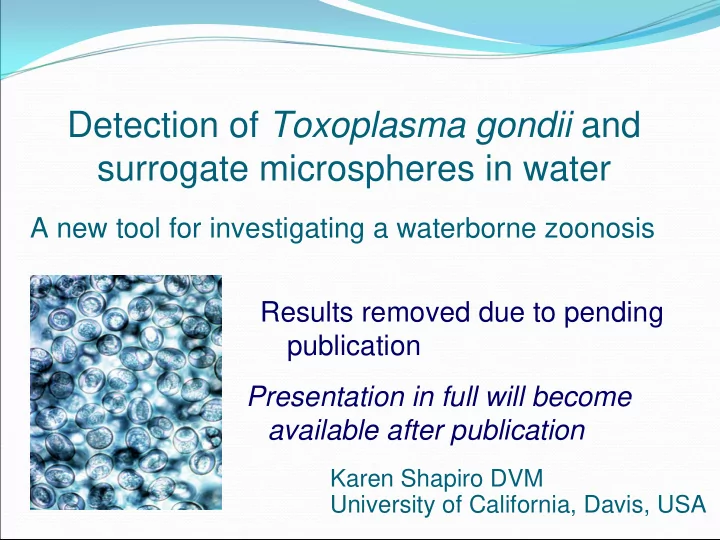
Detection of Toxoplasma gondii and surrogate microspheres in water A new tool for investigating a waterborne zoonosis Results removed due to pending publication Presentation in full will become available after publication Karen Shapiro DVM
Developing nations 3.4 Million deaths annually 1.8 Million children
Viruses Bacteria Parasites
Zoonotic protozoan parasite Infects humans and animals Agent of toxoplasmosis Global distribution Toxo oocyst
Cats definitive hosts Many warm blooded
Oocyst ingestion Undercooked meat Congenital
US: 20% Israel: 40-75% France: 70% Brazil: >90%
David Ferguson, Oxford University
Immunocompromised
during pregnancy
Canada, 1995 Panama, 1979 Brazil, 2002 French Guyana, 1998 India, 2004
Sewage Point source: Storm drains Runoff: Non-point source pollution
Survival in soil 18 mo Survival in water 54 mo
Ultraviolet radiation Radio frequency Bleach Iodine Freezing Desiccation Ozone Ethanol Formalin Salinity
Identify high risk zones
Where do oocysts enter the watershed? Where do oocysts accumulate?
Remove Toxo oocysts
Filtration Coagulation Wetlands
Increased domestic cat
Increased impervious
Storm drains Reduction of natural
Funding: NIH EID Our team:
Veterinary Medicine: Patricia Conrad, Heather Fritz, Jonna Mazet, Ann Melli Environmental Engineering: Stefan Wuertz, Alexander Schriewer Chemistry: Timothy Patten, John Ell, Robert Zasoski Hydrology: Wes Wallender Particle Analysis Laboratory: William Bernt