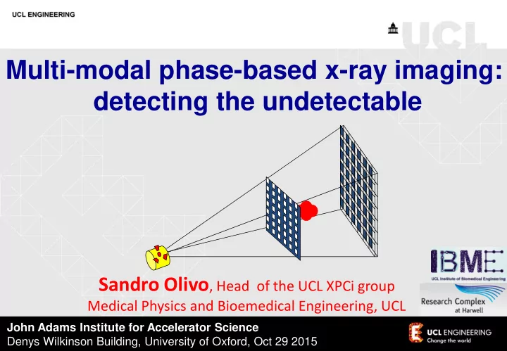

Multi-modal phase-based x-ray imaging: detecting the undetectable Sandro Olivo , Head of the UCL XPCi group Medical Physics and Bioemedical Engineering, UCL John Adams Institute for Accelerator Science Denys Wilkinson Building, University of Oxford, Oct 29 2015
Phase Contrast Imaging vs. Conventional Radiology Refractive index: n = 1 - d i b ; d >> b -> phase contrast ( D I/I 0 ~ 4 pdD z/ l ) >> absorption contrast ( D I/I 0 ~ 4 pbD z/ l Two possible approaches: - detect interference patterns - detect angular deviations
a) absorption b) phase contrast DiMichiel et al Proceedings of MASR1997
Impressive results are achieved in breast imaging absorption phase contrast Arfelli et al. Phys. Med. Biol. 43 (1998) 2845-52
Which led to the realization of a dedicated mammography station in TS Castelli et al. Radiology 259 (2011) 684-94
SRM: findings FFDM obl SRM obl
SRM: findings FFDM obl SRM obl
SRM: findings FFDM obl SRM obl
FSP works wonders when implemented with a spatially coherent source – why ask for more? - It suffer immensely when transferred to conventional sources: the spread associated with projected source size becomes too large and kills the signal. Moreover: The system has little flexibility - only d sd can be changed But: Amazing stuff @ synchrotrons, e.g. check out Cloetens’ work at the ESRF + straightforward use e.g. coupled with Paganin’s single distance phase retrieval Olivo et al. Med. Phys. 28 (2001)1610-19
Other methods to perform phase contrast imaging: “Analyzer Based Imaging” Davis et al, Nature 373 (1995) 595-8; Ingal & Beliaevskaya, J. Phys. D 28 (1995) 2314-7, Chapman et al , Phys. Med. Biol. 42 (1997) 2015-25 - but even before that Forster 1980!
A different way to obtain a similar effect: The Edge Illumination Technique Provides results similar to ABI but opens the way to the use of divergent and polychromatic beams Olivo et al. Med. Phys. 28 (2001)1610-19
THE METHOD CAN BE ADAPTED TO A DIVERGENT AND POLYCHROMATIC (=conventional) SOURCE photons creating increased signal pre-sample apertured mask sample polychromatic, detector divergent detail pixels beam (pre- rotating shaping) anode x-ray source (focal spot 100 m m) photons creating reduced signal detector apertured mask Olivo and Speller Appl. Phys. Lett. 91 (2007) 074106
THE METHOD CAN BE ADAPTED TO A DIVERGENT AND POLYCHROMATIC (=conventional) SOURCE photons creating increased signal pre-sample apertured NB for those of mask sample you who are familiar with grating (or polychromatic, detector divergent Talbot, or detail pixels beam (pre- Talbot-Lau) rotating shaping) interferometers anode x-ray this isn’t one! source (focal spot 100 m m) photons creating reduced signal detector apertured mask Olivo and Speller Appl. Phys. Lett. 91 (2007) 074106
Little loss of signal intensity for source sizes up to 100 µm Which can be achieved with state-of-the-art mammo sources Why? 1) Because we are only relying on refraction, which survives under relaxed coherence conditions; 2) Because we are use aperture pitches matching the pixel size, i.e. BIG: the projected source size remains < pitch, and therefore blurring does “not” occur. Olivo and Speller Phys. Med. Biol. 52 (2007) 6555-73
experimental setup
experimental setup
Preliminary results: the “usual” insects (but a bit faster)
Preliminary results: the “usual” insects (but a bit faster)
Preliminary results: the “usual” insects (but a bit faster)
Preliminary results: the “usual” insects (but a bit faster) Nature 472 (2011) p. 392
Scientific American 305 (2011) p. 14
Preliminary results - mammo (a): GE senographe Essential ADS 54.11; 25 kVp, 26 mAs (b): coded-aperture XPCi, 40 kVp, 25 mA – ENTRANCE dose 7 mGy (< mammo!) It has to be said the tissue was 2.5 cm thick -> we expect ~ same dose for thicker tissues Olivo et al Med. Phys. (letters) 40 (2013) 090701
Preliminary results - mammo (a): GE senographe Essential ADS 54.11; 25 kVp, 26 mAs (b): coded-aperture XPCi, 40 kVp, 25 mA – ENTRANCE dose 7 mGy (< mammo!) It has to be said the tissue was 2.5 cm thick -> we expect ~ same dose for thicker tissues Olivo et al Med. Phys. (letters) 40 (2013) 090701
Preliminary results - mammo (a): GE senographe Essential ADS 54.11; 25 kVp, 26 mAs (b): coded-aperture XPCi, 40 kVp, 25 mA – ENTRANCE dose 7 mGy (< mammo!) Tissue thickness 2 cm-> extrapolation leads to ~standard mammo dose for thicker (4-5 cm) tissues unpublished
Preliminary results - cartilage imaging Rat cartilage, ~ 100 µm thick, invisible to conventional x-rays Marenzana et al, Phys. Med. Biol. 57 (2012) 8173-84 under submission
Cartilage in water: Tells us a lot about CAXPCi vs Talbot/Lau sensitivity: TALBOT/LAU CAXPCI (PORK cartilage) (RAT cartilage) from Stutman et al , Phys. Med. Biol. 56 (2011) 5697-720 - SAME signal (despite thicker cartilage for T/L); - on 3 rd Talbot order (cartilage in water invisible on 1 st order) Marenzana et al, Phys. Med. Biol. 57 (2012) 8173-84
More on the sensitivity of the lab system: 6 1.0 µrad (a) (b) 6 2 0.5 µrad (a) (b) (c) (d) (e) (f) µrad µrad 0.0 -0.5 4 -1.0 0 0 1 2 2 -6 -2 0 1.0 5 (g) (h) 4 0.8 Theory Theory 3 Retrieved 0.6 Refraction angle (µrad) Refraction angle (µrad) Retrieved 2 0.4 -2 0.2 1 2 mm 0 0.0 0 200 400 600 0 200 400 600 -1 -0.2 0.6 -2 -0.4 0.4 0.2 -0.6 -3 0.0 -4 -0.2 -0.8 -4 -0.4 Position (µm) Position (µm) -5 -0.6 -1.0 1 mm -6 This gives a phase sensitivity of ~ 270 nRad, with only 2 images x 7s exposure each; same as reported by Thuring (Stampanoni’s group) for GI. Revol reported a sensitivity of about 110 nRad but with 12 x 7s frames – as one can expect the value to scale with sqrt(exp time), that also fits. Diemoz et al , Appl. Phys. Lett. 103 (2013) 244104
Actually we’ve done much better on cartilage, in collaboration with PIXIRAD (Bellazzini et al. ) 26 kVp Underpins sensitivity better than 270 nrad – indeed we’ve recently measured 150-200 and are putting measures in place to improve it even further. Endrizzi et al , JINST 9 (2014) C11004
Quantitative phase contrast imaging “ SLOPE - ” “ SLOPE + ” Titanium Aluminum PEEK Highly precise retrieval, for both high and low Z materials, up to high gradients where other methods break down Munro et al Opt. Exp. 21 (2013) 647-61
Phase retrieval with synchrotron and conventional sources: Ti filament: retrieved @ synchrotron and with conventional source! @ conventional source: incoherence modelled as beam spreading – the movement of the “spread” beam is then tracked and referred back to the phase shift that caused it. But with lots of care as far as “effective energy” is concerned! (See Munro & Olivo Phys. Rev. A 87 (2013) 053838) Munro et al , PNAS 109 (2012) 13922-7
preliminary CT results Soft tissue inside wasp thorax resolved Dose tens of mGy , instead of tens of Gy! Hagen et al, Med. Phys. (letters) 41 (2014) 070701
preliminary CT results AVAILABLE ONLINE—See http://www.medphys.org July 2014 Volume 41, Number 7 The International Journal of Medical Physics Research and Practice Soft tissue inside wasp thorax resolved Dose tens of mGy , instead of tens of Gy! First experimentally acquired x-ray phase-contrast images acquired with ordinary x-ray source using edge-illumination method (EI PCi). (1) 3D schematic view of the laboratory implementation of tomographic EI XPCi. (a) Views from top showing two opposing edge illumination conditions, (b,c), achie ved by shifting the sample mask appropriately. (2) Coronal tomographic images of a w asp showing the phase shift (a) and attenuation (b) images within the insect with profi les extracted across the indicated thorax region. (3) 3D volume rendering of the wasp derived from phase shift images. [Figures 1, 2, and 3 from Hagen, Munro, Endrizzi, Diemoz, and Oli v o, “Lo w-dose phase contrast tomography with conventional x-ray sources, ” Med. Phys. 41, 070701 (5pp.) (2014)]. Published by the American Association of Physicists in Medicine (AAPM) with the association of the Canadian Organization of Medical Physicists (COMP), the Canadian College of Physicists in Medicine (CCPM), and the International Organization for Medical Physics (IOMP) through the AIP Publishing LLC. Medical Physics is an of fi cial science journal of the AAPM and of the COMP/CCPM/IOMP. Medical Physics is a hybrid gold open-access journal. Hagen et al, Med. Phys. (letters) 41 (2014) 070701
preliminary CT results Rabbit oesophagous Hagen et al , submitted to Sci. Rep.
Three-shot DARK FIELD IMAGING retrieval Endrizzi et al , Appl. Phys. Lett. 104 (2014) 024106
DARK FIELD IMAGING of breast calcifications 3 images only, still within clinical dose limits! ENTRANCE dose 12 mGy (still compatible with mammo) Endrizzi et al , Appl. Phys. Lett. 104 (2014) 024106
Non-medical applications: testing of composite materials Endrizzi et al , Compos. Struct. 134 (2015) 895-9
Microbubbles: a new concept of “phase - based” x -ray contrast agent Millard et al. Appl. Phys. Lett. 103 (2013) 114105
Recommend
More recommend