Correction of Dentofacial Deformities (Orthognathic Surgery) Dr. - - PDF document

correction of dentofacial deformities
SMART_READER_LITE
LIVE PREVIEW

Correction of Dentofacial Deformities (Orthognathic Surgery) Dr. - - PDF document

Correction of Dentofacial Deformities (Orthognathic Surgery) Dr. Rafik Al Kowafi BDS, MSc, German board of Oral and Maxillofacial Surgery ( Berlin-Germany), Doctoral degree by LBMS Definition Orthognathic surgery is a combination of


slide-1
SLIDE 1

1

Correction of Dentofacial Deformities

(Orthognathic Surgery)

  • Dr. Rafik Al Kowafi BDS, MSc, German board of Oral and

Maxillofacial Surgery ( Berlin-Germany), Doctoral degree by LBMS

Definition

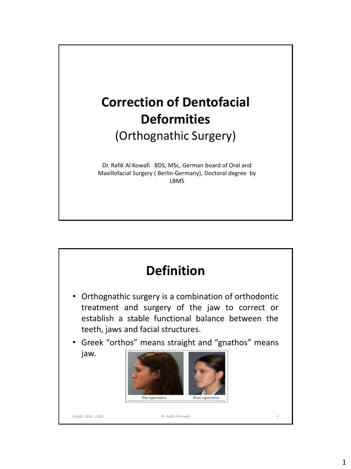
  • Orthognathic surgery is a combination of orthodontic

treatment and surgery of the jaw to correct or establish a stable functional balance between the teeth, jaws and facial structures.

  • Greek “orthos” means straight and “gnathos” means

jaw.

2

  • Dr. Rafik Al Kowafi

4 April 2016 LIMU

slide-2
SLIDE 2

2

Aims of orthognathic surgery

  • To treat any jaw imbalance and the resulting

incorrect bite, which could adversely affect the cosmetic (esthetic) appearance as well as the proper functioning of the teeth.

  • Aims:

1.Function: Normal chewing, speech, respiratory function. 2.Esthetics: Establish facial harmony and balance. 3.Stability: Avoid short and long term relapse. 4.Minimize orthodontic treatment time.

  • Dr. Rafik Al Kowafi

3 4 April 2016 LIMU

Causes of dentofacial deformity

1. Congenital (e.g, hemifacial microsomia, mandibulofacial dysostosis “Treacher-Collins syndrome”, cleft lip and palat). 2. Prenatal Problems (e.g. hypoplasia of midface due to fetal alcohol syndrome). 3. Environmental influences (e.g. abnormal tongue and lip postures, mouth breathing). 4. Trauma (e.g. TMJ trauma).

4

  • Dr. Rafik Al Kowafi

4 April 2016 LIMU

slide-3
SLIDE 3

3

Causes of dentofacial deformity

  • Dr. Rafik Al Kowafi

5

Treacher Collins syndrome Hemifacial Microsomia TMJ trauma

4 April 2016 LIMU

Maxillofacial deformities

(1) Dental dysplasias (2) Skeletal dysplasias (3) Dento-Skeletal dysplasias

6

  • Dr. Rafik Al Kowafi

4 April 2016 LIMU

slide-4
SLIDE 4

4

Dental Dysplasias

  • Dental

dysplasias are limited strictly to malocclusions that result from abnormal relationship of the dentition and not from the skeletal position of the upper and lower jaws.

  • These can be corrected with orthodontic

treatment.

A- Normal occlusion B- Dental Dysplasias C- Skeletal Dysplasias (Maxilla) D- Skeletal Dysplasias (Mandible)

7

  • Dr. Rafik Al Kowafi

4 April 2016 LIMU

Skeletal Dysplasias

  • In patients with skeletal dysplasia only, the

dentition is in good alignment, but the maxilla and/or mandible are dysplastic

  • Skeletal

dysplasias require correcting the skeletal deformity without altering the

  • cclusion.

8

  • Dr. Rafik Al Kowafi

4 April 2016 LIMU

slide-5
SLIDE 5

5

Dento-Skeletal Dysplasias

  • In dento-skeletal

dysplasias, the dentition is mal-positioned within each arch and with each

  • ther; additionally, the

skeletal relationship of the upper and lower jaws is abnormal.

  • These are corrected with
  • rthognathic surgery

( ortho + surgery)

9

  • Dr. Rafik Al Kowafi

4 April 2016 LIMU 10

Clinical examination: 1- full face and profile 2- photographic document 3- dental arch examination 4- TMJ and muscles of mastication 5- Steriolithic model Radiographic examination; 1. Cephalometric 2. Panoramic 3. Postero-anterior facial films 4. T.M.J. films 5. CBCT 6. C.T. scan

  • Dr. Rafik Al Kowafi

Evaluation of patients with dentofacial deformities

Steriolithic model

4 April 2016 LIMU

slide-6
SLIDE 6

6

Evaluation of patients with dentofacial deformities

4 April 2016 LIMU

  • Dr. Rafik Al Kowafi

11

A, Cone beam computed tomography scan clearly demonstrating bone deformity in three dimensions. B, Stereolith graphic model.

A B

Evaluation of patients with dentofacial deformities

4 April 2016 LIMU

  • Dr. Rafik Al Kowafi

12

Model surgery used to determine direction and distance of surgical movement necessary to achieve desired postoperative

  • cclusion and

facial esthetics.

slide-7
SLIDE 7

7

Evaluation of patients with dentofacial deformities

4 April 2016 LIMU

  • Dr. Rafik Al Kowafi

13

A and B, CT and CBCT Three-dimensional imaging and virtual planning. C and D, The splints are then designed and constructed using CAD-CAM rapid prototyping technology.

Treatment Phases

1-PRESURCICAL TREATMENT PHASE

  • Periodontal Considerations
  • Restorative Considerations
  • Presurgical Orthodontic Considerations
  • Final Treatment Planning

2- SURGICAL TREATMENT PHASE 3- POSTSURGICAL TREATMENT PHASE

  • Completion of Orthodontics
  • Postsurgical Restorative and Prosthetic Considerations
  • Dr. Rafik Al Kowafi

14 4 April 2016 LIMU

slide-8
SLIDE 8

8

Timing of Surgery

  • Usually done when all growth is complete.
  • Assessed

by superimposition

  • f

serial lateral cephalometrics.

  • Can be performed when growth is not yet complete

in cases of psychosocial problems or great severity when function is compromised (i.e. breathing, chewing).

15

  • Dr. Rafik Al Kowafi

4 April 2016 LIMU

Procedures

  • The surgery might involve one jaw, or the two jaws at

the same time (bi-maxillary osteotomy). Steps:

  • Making cuts as planned in the bones (osteotomy).
  • Repositioning the cut pieces in the desired alignment.
  • Fixation by wires, plates, or screws.

Methods of bone cutting:

  • By using special electrical saws, burs and manual

chisels.

  • Recently by using ultra-sound waves (piezo surgery).

16

  • Dr. Rafik Al Kowafi

4 April 2016 LIMU

slide-9
SLIDE 9

9

Types of Dentofacial abnormalities

  • 1. Mandibular Excess
  • 2. Mandibular Deficiency
  • 3. Maxillary Excess
  • 4. Maxillary and Midface Deficiency
  • 5. Combination Deformities and Asymmetries
  • Dr. Rafik Al Kowafi

17 4 April 2016 LIMU

Mandibular Excess

(Mandibular Prognathism)

Clinical features:

  • Abnormal
  • cclusion

with class III molar and cuspid relationships

  • A

reverse

  • verjet

in the incisor area with posterior cross bite

  • Flat appearance of the mid

face

  • Concave profile
  • Obtuse gonial angle.
  • Diminished labio-mental fold
  • Acute naso-labial angle
  • Posterior cross bite
  • Dr. Rafik Al Kowafi

18 4 April 2016 LIMU

slide-10
SLIDE 10

10

Surgical techniques for correction of mandibular prognathism

1) Bilateral sagittal split osteotomy (BSSO) 2) Vertical ramus osteotomy 3) Body osteotomy 4) Anterior mandibular subapical osteotomy.

  • Dr. Rafik Al Kowafi

19 4 April 2016 LIMU

1-Bilateral sagittal split osteotomy (BSSO)

  • The osteotomy splits

the ramus and posterior body of the mandible in a sagittal fashion which allows either setback

  • r

advancement of the mandible.

  • Dr. Rafik Al Kowafi

20 4 April 2016 LIMU

slide-11
SLIDE 11

11

2-Vertical ramus osteotomy

  • In this technique the lateral aspect of the ramus is

exposed through a submandibular incision, the ramus is sectioned in a vertical fashion, and the entire body and anterior ramus section of the mandible are moved posteriorly, which places the teeth in proper occlusion.

4 April 2016 LIMU

  • Dr. Rafik Al Kowafi

21

2-Vertical ramus osteotomy

4 April 2016 LIMU

  • Dr. Rafik Al Kowafi

22

slide-12
SLIDE 12

12

3- Body osteotomy

  • By removing sections of bone in the body of

the mandible, which allowed the anterior segment to be moved posteriorly.

  • Could be done through intra-oral, or extra-oral

approach or combination.

4 April 2016 LIMU

  • Dr. Rafik Al Kowafi

23

4- Anterior mandibular subapical

  • steotomy.
  • When

the reverse

  • verjet

relationship is isolated to the anterior area of the mandible, a subapical osteotomy technique can be used for correction of mandibular dental prognathism

  • In

this technique, bone is removed in the area

  • f

an extraction site of a bicuspid or molar tooth, and the anterior dentoalveolar segment of the mandible is moved to a more posterior position.

  • Dr. Rafik Al Kowafi

24 4 April 2016 LIMU

slide-13
SLIDE 13

13

Mandibular Deficiency

(Mandibular Retrusion or Micrognathia)

Clinical features:

  • Retruded position of the chin as viewed from the profile (bird face deformity)
  • Excess labio-mental fold
  • Abnormal posture of the upper lip, and poor throat form.
  • Intraorally, class II molar and cuspid relationships
  • An increased overjet in the incisor area with Incisor crowding in the lower jaw
  • Acute gonial angle.

4 April 2016 LIMU

  • Dr. Rafik Al Kowafi

25

Surgical techniques for correction of mandibular deficiency

1) Vertical osteotomy and iliac crest bone grafts in the osteotomy defect. 2) Bilateral sagittal split osteotomy (BSSO) 3) Total mandibular subapical osteotomy. 4) Inferior border osteotomy (Genioplasty) with advancement.

  • Dr. Rafik Al Kowafi

26 4 April 2016 LIMU

slide-14
SLIDE 14

14 1- Vertical osteotomy and iliac crest bone grafts in the

  • steotomy defect.
  • Mandibular advancement using

vertical osteotomy and iliac crest bone grafts in osteotomy defect.

  • Dr. Rafik Al Kowafi

27

  • This procedure is easily accomplished

through an intraoral incision, The significant bony overlap produced with the BSSO allows for adequate bone healing and improved postoperative stability.

2-Bilateral sagittal split osteotomy (BSSO)

4 April 2016 LIMU

Preoperative facial esthetics demonstrating clinical features of mandibular deficiency Preoperative occlusion demonstrating Class II relationship and overjet

4 April 2016 LIMU

  • Dr. Rafik Al Kowafi

28

slide-15
SLIDE 15

15

Bilateral sagittal split osteotomy with advancement of mandible.

4 April 2016 LIMU

  • Dr. Rafik Al Kowafi

29

3- Total mandibular subapical

  • steotomy:
  • Indicated

when the antero-posterior position of the chin is adequate but a class II malocclusion exists.

  • By combining the osteotomy with

interpositioned bone grafts, this technique can he used to increase lower facial height.

4- Inferior border osteotomy (Genioplasty):

  • When a proper occlusal relationship

exists or when anterior positioning of the mandible would not be sufficient to produce adequate projection of the chin, an inferior border osteotomy (i.e., genioplasty) with advancement is performed.

  • Dr. Rafik Al Kowafi

30 4 April 2016 LIMU

slide-16
SLIDE 16

16

Maxillary excess (maxillary protrusion)

Vertical, transverse and anteroposterior

  • Clinical features:

– Elongation of the lower third

  • f the face

– Excessive gingival & incisal exposure (gummy smile). – Lip incompetence – A narrow nose – Convex facial profile – Class II molar occlusion – High arched palate.

  • Dr. Rafik Al Kowafi

31 4 April 2016 LIMU

Maxillary and Midface Deficiency

  • Clinical features:

– A retruded upper lip. – Deficiency of the paranasal and infraorbital rim areas. – Inadequate tooth exposure during smile. – A prominent chin relative to the middle third of the face – A class III malocclusion with reverse anterior overjet

32

  • Dr. Rafik Al Kowafi

4 April 2016 LIMU

slide-17
SLIDE 17

17

Orthognathic surgeries of maxilla and midface

1. Anterior maxillary osteotomy ( Wassmund approach).

  • 2. Total maxillary osteotomy.

– Le fort I osteotomy. – Le fort II osteotomy. – Lefort III osteotomy.

33

  • Dr. Rafik Al Kowafi

4 April 2016 LIMU

Anterior maxillary osteotomy (wassmund 1935)

  • Indicated for correction of dento-alveolar

protrusion of anterior maxilla.

  • The anterior segment can be moved:

Superiorly, Inferiorly, posteriorly.

Indications:

  • maxillary protrusion
  • marked protrusion of

maxillary teeth

  • open bite

34

  • Dr. Rafik Al Kowafi

4 April 2016 LIMU

slide-18
SLIDE 18

18

Total maxillary osteotomy.

(complete le fort I osteotomy)

  • By creating a le fort I fracture to

allow mobilization of the maxilla and articulation in any other position desired. ( backward/ forward’ upward’ downward/ rotation), Then fixation in its new place using miniplates. Indications:

1. Treatment

  • f

maxillary protrusion and retrusion. 2. Correction of open and closed bite.

35

  • Dr. Rafik Al Kowafi

4 April 2016 LIMU

Le Fort II osteotomy

  • For patients with central

midface hypoplasia extending into the naso-ethmoidal area.

  • It allows a certain amount of

lengthening of the midface, especially of the nose with a complete advancement of the central midface.

36

  • Dr. Rafik Al Kowafi

4 April 2016 LIMU

slide-19
SLIDE 19

19

  • Complete craniofacial dysjunction

by the LeFort III osteotomy allows the surgeon to alter the orbital position and volume, zygomatic projection, position of the nasal root, fronto-nasal angle, and position of the maxilla and to lengthen the nose. used primarily for correction of total midface

hypoplasia, usually

  • f

cranio- synostotic

  • rigin

as in: Alpert syndrome and Crouzon's syndrome.

Le Fort III osteotomy

37

  • Dr. Rafik Al Kowafi

4 April 2016 LIMU

Le Fort III osteotomy

4 April 2016 LIMU

  • Dr. Rafik Al Kowafi

38

slide-20
SLIDE 20

20

bi-maxillary osteotomy

( Maxillary and mandibular osteotomy)

Le Fort I osteotomy for maxillary advancement and bilateral sagittal

  • steotomies for setback of the mandible.

4 April 2016 LIMU

  • Dr. Rafik Al Kowafi

39

bi-maxillary osteotomy

( Maxillary and mandibular osteotomy)

Postoperative facial appearance.

4 April 2016 LIMU

  • Dr. Rafik Al Kowafi

40

slide-21
SLIDE 21

21

bi-maxillary osteotomy

( Maxillary and mandibular osteotomy)

Postoperative occlusion and postoperative radiograph

4 April 2016 LIMU

  • Dr. Rafik Al Kowafi

41

Complications of orthognathic surgery

  • 1. Vascular complications.
  • 2. Nonunion or delayed union.
  • 3. Dental and periodontal defects.
  • 4. Nerve injuries.
  • 5. Unanticipated fractures.
  • 6. Temporomandibular joint dysfunction.
  • 7. Postoperative occlusal discrepancies.
  • 8. Facial scars.

42

  • Dr. Rafik Al Kowafi

4 April 2016 LIMU

slide-22
SLIDE 22

22

Distraction Osteogenesis

 Distraction osteogenesis (DO), also called callus distraction, callotasis and osteodistraction  It is the process of generating new bone in a gap, created by

  • steotomy, between two bone

segments in response to the application of graduated controlled tensile stress across the gap

 It relies on the normal healing capacity of the own body that

  • ccurs between the surgically
  • steotomized bone segments

43

  • Dr. Rafik Al Kowafi

4 April 2016 LIMU

Advantages of DO

  • 1. Large volume of new bone formation.
  • 2. Simultaneous regeneration of both hard and soft

tissues.

  • 3. Complex 3D bone reconstruction.
  • 4. No additional bone graft.
  • 5. Decreased bone resorption.
  • 6. Less invasive.
  • 7. Less relapse.

44

  • Dr. Rafik Al Kowafi

4 April 2016 LIMU

slide-23
SLIDE 23

23

Indications of distraction Osteogenesis

1. Children or infants with severe retrognathia associated with a syndrome (Pierre Robin syndrome, Treacher Collins). 2. Unilateral hypoplasia of the mandible (Hemifacial microsomia). 3. Mandibular hypoplasia due to trauma and/or ankylosis of the temporomandibular joint. 4. Nonsyndromic mandibular hypoplasia associated with a dental malocclusion where movement of mandible required is >10mm 5. Severe obstructive sleep apnea in patients who are morbidly

  • bese.
  • 6. Shortened vertical height of the alveolar bone to receive an

implant

4 April 2016 LIMU 45

  • Dr. Rafik Al Kowafi

Distraction Osteogenesis

  • Craniofacial distraction devices classification:

1. External (bone borne) 2. Internal (tooth borne or bone borne).

a. Intraoral. b. Subcutaneous.

  • According to the direction of bone formation

can be classified as:

  • 1. Unidirectional
  • 2. Bidirectional
  • 3. Multidirectional

4 April 2016 LIMU

  • Dr. Rafik Al Kowafi

46

slide-24
SLIDE 24

24

Distraction Osteogenesis

Unidirectional Intraoral device

4 April 2016 LIMU 47

  • Dr. Rafik Al Kowafi

Distraction Osteogenesis

4 April 2016 LIMU

  • Dr. Rafik Al Kowafi

48

slide-25
SLIDE 25

25

Distraction Osteogenesis

4 April 2016 LIMU

  • Dr. Rafik Al Kowafi

49

Distraction Osteogenesis

4 April 2016 LIMU

  • Dr. Rafik Al Kowafi

50

slide-26
SLIDE 26

26

Distraction Osteogenesis

Unidirectional extraoral device

4 April 2016 LIMU 51

  • Dr. Rafik Al Kowafi

Distraction Osteogenesis

Bidirectional extraoral device

4 April 2016 LIMU 52

  • Dr. Rafik Al Kowafi
slide-27
SLIDE 27

27

Distraction Osteogenesis

Multidirectional extraoral device

4 April 2016 LIMU 53

  • Dr. Rafik Al Kowafi

Distraction Osteogenesis

Sequential periods of DO:

  • 1. Osteotomy.
  • 2. Latency.
  • 3. Distraction.
  • 4. Consolidation.
  • 5. Remodeling.

4 April 2016 LIMU 54

  • Dr. Rafik Al Kowafi
slide-28
SLIDE 28

28

1- Osteotomy

  • Division of bone in two segments.
  • Triggers bone healing ( # healing )
  • Recruitment of osteoprogenitor cells.
  • Osteoinduction
  • Osteoconduction
  • Typically, a linear osteotomy is created through the

mandible with burs or saws, except in the location of the inferior alveolar neurovascular bundle. The osteotomy is completed with osteotomes

4 April 2016 LIMU 55

  • Dr. Rafik Al Kowafi

2- Latency period

  • Period from bone division to onset of traction.
  • Represents time allowed for callus formation.
  • Sequence of events

– Hematoma – Clot – Bone necrosis at the ends of # segments – Ingrowths of vasoformative elements & cellular proliferation – Stage of inflammation ( 1-3 days ) – Clot is replaced by granulation tissue – Granulation tissue is converted to fibrous and cartilage tissue – callus formation

  • The latency period ranges from 0 to 10 days, although the

most common latency period is 5 days, and is applicable in adults.

4 April 2016 LIMU 56

  • Dr. Rafik Al Kowafi
slide-29
SLIDE 29

29

3- Distraction period

  • Application of traction forces to osteotomised bone

segments.

  • Bone segments are gradually pulled apart resulting in

formation of new bone tissue with in progressively increasing inter-segmentary gap.

  • The gold standard for clinical distraction osteogenesis is

1 mm per day, divided into 2 or 4 activations per day.

4 April 2016 LIMU 57

  • Dr. Rafik Al Kowafi

4- Consolidation period

  • Time between cessation of traction and removal of

distraction devices.

  • This period represents the time required for complete

mineralization.

  • The consolidation period in adults should be a minimum
  • f 3 months and can extend up to 6 months as needed.
  • Consolidation time is related to the magnitude of the

distraction distance and the age of the patient.

4 April 2016 LIMU 58

  • Dr. Rafik Al Kowafi
slide-30
SLIDE 30

30

5- Remodeling period

Period from the application of full functional loading to the complete remodeling of newly formed bone.

Last stage of cortical reconstruction ( 1 year )

4 April 2016 LIMU 59

  • Dr. Rafik Al Kowafi