
SLIDE 1
2019/10/25 1
A Pot-Pourri of Pitfalls in Non-GYN Cytopathology
Cady Zeman-Pocrnich October 26, 2019
Conflict of Interest Disclosure
- Member of the IQMH Cytopathology Scientific
Committee
Objectives
- After this session on Non-GYN pitfalls,
participants should be able to:
- Appropriately classify lesions from a variety of
Non-GYN sites by correctly applying morphological criteria, ancillary study criteria, and clues from the clinical history;
- Reflect on diagnostic misses and near misses in
Non-GYN cytopathology
Outline
Pitfalls I have encountered in the cytopathological diagnosis of:
- Neuroendocrine Lesions
- Salivary Gland Tumours
- Thyroid Nodules
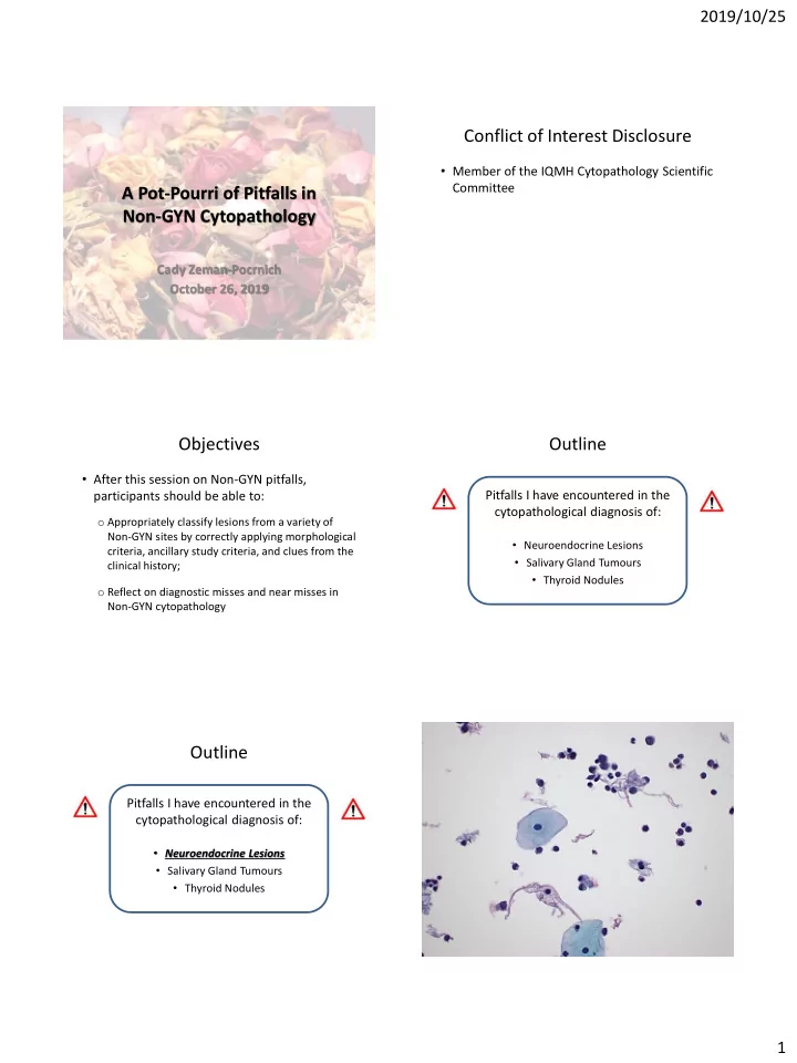
Outline
Pitfalls I have encountered in the cytopathological diagnosis of:
- Neuroendocrine Lesions
- Salivary Gland Tumours
- Thyroid Nodules