
SLIDE 1
3/16/2015 1
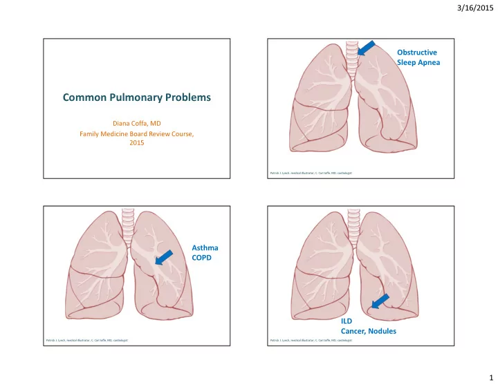
Common Pulmonary Problems
Diana Coffa, MD Family Medicine Board Review Course, 2015
Patrick J. Lynch, medical illustrator; C. Carl Jaffe, MD, cardiologist
Obstructive Sleep Apnea
Patrick J. Lynch, medical illustrator; C. Carl Jaffe, MD, cardiologist
Asthma COPD
Patrick J. Lynch, medical illustrator; C. Carl Jaffe, MD, cardiologist