
SLIDE 1
1
Clostridium perfringens
Dean O. Cliver (materials from M. N. Hajmeer)
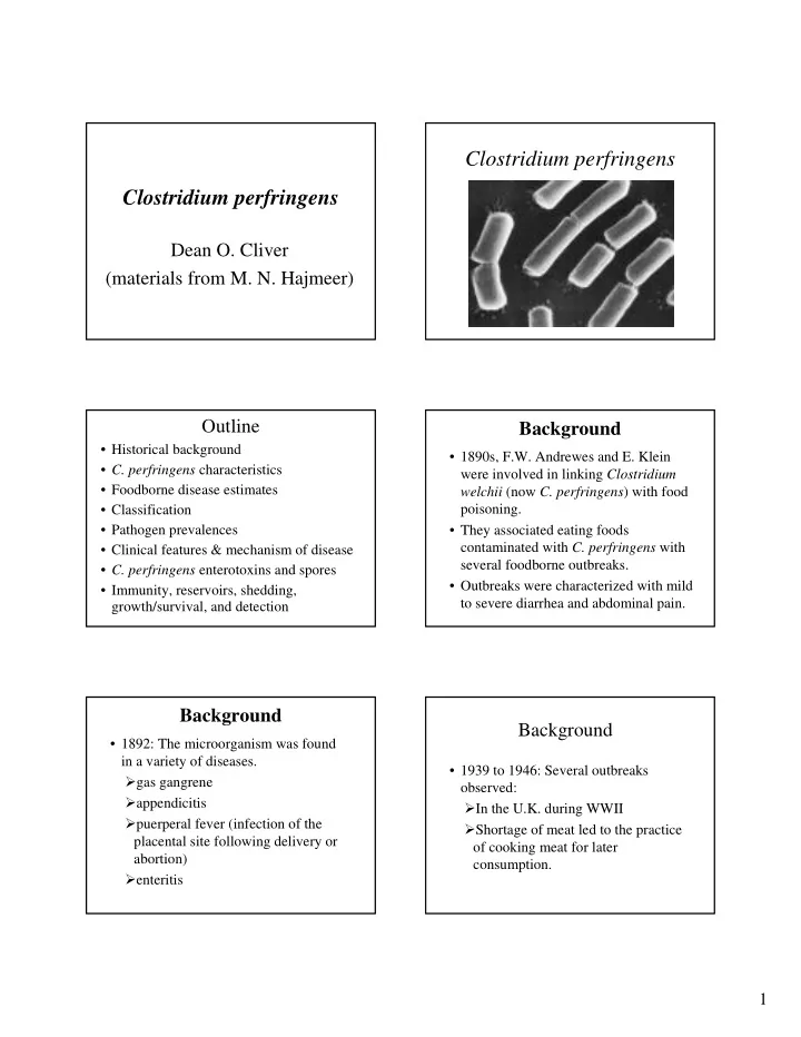
Clostridium perfringens
Outline
- Historical background
- C. perfringens characteristics
- Foodborne disease estimates
- Classification
- Pathogen prevalences
- Clinical features & mechanism of disease
- C. perfringens enterotoxins and spores
- Immunity, reservoirs, shedding,
growth/survival, and detection
Background
- 1890s, F.W. Andrewes and E. Klein
were involved in linking Clostridium welchii (now C. perfringens) with food poisoning.
- They associated eating foods
contaminated with C. perfringens with several foodborne outbreaks.
- Outbreaks were characterized with mild
to severe diarrhea and abdominal pain.
Background
- 1892: The microorganism was found
in a variety of diseases. gas gangrene appendicitis puerperal fever (infection of the placental site following delivery or abortion) enteritis
Background
- 1939 to 1946: Several outbreaks
- bserved:
In the U.K. during WWII Shortage of meat led to the practice
- f cooking meat for later