SLIDE 1 1
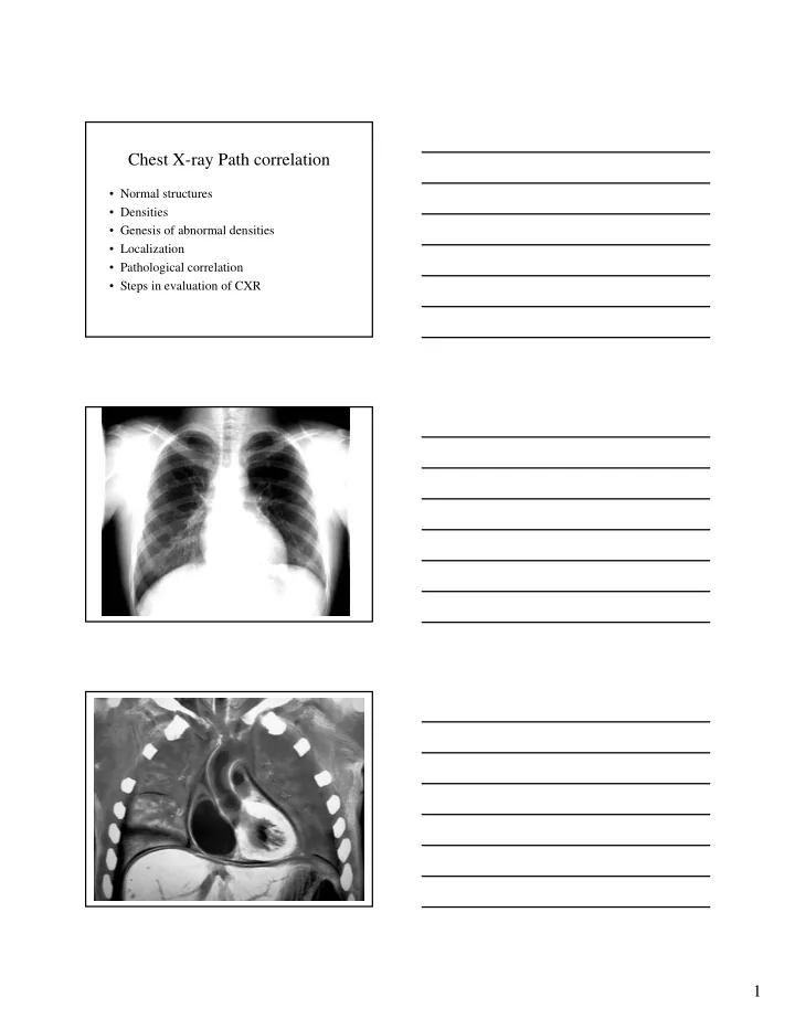
Chest X-ray Path correlation
- Normal structures
- Densities
- Genesis of abnormal densities
- Localization
- Pathological correlation
- Steps in evaluation of CXR
SLIDE 2
2
Left RA RV LV LA
SLIDE 3
3
SLIDE 4
4
Densities
SLIDE 5 5
H e m Hemorrhage
Localization
- Lobar distribution
- Air bronchogram
- Silouhette sign
- Extra pleural sign
SLIDE 6
6
SLIDE 7
7
Lobar density Silouhette sign
SLIDE 8
8
Silouhette sign
SLIDE 9
9
RLL LLL Lingula RML LUL RUL Silouhette sign Extrapleural sign
SLIDE 10
10
Extrapleural sign Peripheral Sharp inner Indistinct outer Concave angles Air bronchogram
SLIDE 11 11
Pathological correlation
- Consolidation
- Cavitation
- Mass
- Atelectasis
- Pleural effusion
- Pneumothorax
- Pulmonary edema
Consolidation Pneumonia
SLIDE 12
12
Air bronchogram Lobar No loss of volume Consolidation Cavity Hole Wall Lumen Content
SLIDE 13
13
Mass Mass Homogenous Sharp margin No respect Atelectasis
SLIDE 14
14
Atelectasis Loss of lung volume Pleural effusion Homogenous Costophrenic angle Meniscus
SLIDE 15
15
Pneumothorax Pneumothorax Air in pleura Atelectatic lung Hemithorax Mediastinual shift Pulmonary edema
SLIDE 16
16
Pulmonary edema Pulmonary edema Bilateral Diffuse Alveolar Emphysema
SLIDE 17
17
Emphysema Bleb Bullous emphysema Emphysema
SLIDE 18
18
Flat diaphragm Emphysema Emphysema Dark lungs AP diameter Low flat diaphragms Vertical heart Retrosternal air Infracardiac air Avascularity Blebs
SLIDE 19
19
Mediastinal nodes Metastases Honeycombing
SLIDE 20
20
Interstitial fibrosis Diffuse interstitial Diffuse interstitial Lines Honeycombing Nodules
SLIDE 21 21
Steps in evaluation of CXR
- Identify abnormalities
- Localize
- Identify pathological process
- Probable etiology
- Tests to confirm
Case 1 Case 2
SLIDE 22
22
Case 3 Case 4
SLIDE 23
23
Case 5
SLIDE 24
24
Case 6 Case 7 Case 8