Chemistry Review Case Study #1 A 42-year old male presents with - - PowerPoint PPT Presentation

chemistry review case study 1
SMART_READER_LITE
LIVE PREVIEW

Chemistry Review Case Study #1 A 42-year old male presents with - - PowerPoint PPT Presentation

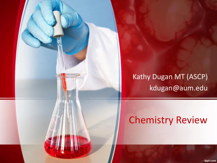
Kathy Dugan MT (ASCP) kdugan@aum.edu Chemistry Review Case Study #1 A 42-year old male presents with anorexia, nausea, fever and icterus of the skin and mucous membranes. He noticed that his urine had appeared dark for the past several


slide-1
SLIDE 1

Chemistry Review

Kathy Dugan MT (ASCP) kdugan@aum.edu

slide-2
SLIDE 2

Case Study #1

  • A 42-year old male presents with anorexia, nausea, fever and icterus of

the skin and mucous membranes. He noticed that his urine had appeared dark for the past several days. The physician orders a series of tests. Based on the following test results, what is the most likely diagnosis? – ALP: 240 U/L (36-92 U/L) AST: 1500 U/L (0-35 U/L) – GGT: 75 U/L (0-30 U/L) Total bilirubin: 1.9 mg/dL (0.-1.2 gm/dL) – Urine bilirubin: positive Fecal urobilinogen: decreased – A. Acute hepatitis – B. Alcoholic cirrhosis – C. Metastatic carcinoma of the pancreas – D. Obstructive jaundice

slide-3
SLIDE 3
  • AST (0-35 U/L)

– will be very elevated in liver disease; Maybe 100 x normal with viral hepatitis – Used to diagnose and monitor hepatocellular disease

  • GGT (0-30 U/L)

– Very elevated in obstructive disease (hepatobilliary) – Moderate increase in hepatocellular – ALP + GGT are increased = Liver NOT bone

  • ALP ( 36-92 U/L)

– Moderate Increase (3x normal) with extra hepatobiliary disease – Biliary obstruction causes synthesis of ALP so will be very increased

  • Bilirubin (Total 0.3-1.2 mg/dL; direct 0-0.3 mg/dL)

– Must pass through liver to be conjugated (water soluble)= direct bilirubin – Direct  bile duct  gall bladder  duodenum  bacteria reduce to urobilinogen  excreted in feces

  • Urobilinogen (Fecal)

– Decreased (chalk-clay color to feces) amounts indicate liver obstruction or liver disease

slide-4
SLIDE 4

Case Study # 2

  • A 4-year old male child is brought to the pediatrician because

the parents are concerned about the child’s frequent falling, which results in bruising. The parents indicate that the child has difficulty running, walking, standing up, climbing stairs, and even sitting up straight. The child also appears somewhat

  • weak. Which enzyme(s) would you expect to be elevated?

– A. AST

  • B. ALP

– C. LD

  • D. CK

– What diagnosis would you expect?

  • Duchene's Muscular Dystrophy
slide-5
SLIDE 5
  • Duchene’s Muscular Dystrophy

– Muscle weakness due to destruction of muscle fibers – X-linked recessive disorder, symptoms start in males 3-7 yrs old – CK

  • 50-100X normal. Indicates muscle destruction

– AST & LD

  • Increased because they are found in skeletal muscle

– ALP

  • Not present in skeletal muscle
  • Used to assess hepatobiliary and bone disorders
slide-6
SLIDE 6

Case Study #3

  • A healthy active 10-year old boy with no prior history of illness

comes to the lab after school for a routine chemistry screen in

  • rder to meet requirements for summer camp. After

centrifugation, the serum looks cloudy. The specimen had the following results – Blood glucose: 135 mg/dL (70-100 mg/dL) – Total cholesterol: 195 mg/dL ((150-199 mg/dL) – Triglyceride: 185 mg/dL (<250 mg/dL)

  • What would be the most probable explanation

– A. Risk for coronary artery disease – B. Has Type 1 Diabetes mellitus that is undiagnosed – C. Has an inherited genetic disease causing lipid imbalance – D. Was most likely not fasting when the specimen was drawn

slide-7
SLIDE 7
  • This is a 10 year-old healthy child. He is probably not suffering from

any lipid disorder or glucose disorder. – Lipids were within high normal range – Glucose was elevated, but if from a non-fasting (random) specimen was within an acceptable range

  • Should never be >200 mg/dL
  • Since he came directly from school, he was probably not fasting.
slide-8
SLIDE 8

Case Study #4

  • A 68-year old male in an unconscious state is transported to the

emergency department after being involved in a one-car crash, where he drove off the road and hit a tree. Because he was alone at the time and there was no apparent cause for the accident, it is assumed that he blacked out, which caused him to lose control of the car. He was not wearing a seat belt and has a broken leg, multiple contusions, and cuts. Blood samples were drawn upon arrival to the ED and in 3-hr intervals for 12 hours; all control values were within acceptable range. Selected test results follow:

slide-9
SLIDE 9

Test Initial Values 3 Hours 9 Hours Reference Ranges Myoglobin 57 ng/mL 140 ng/mL 281 ng/mL 30-90 ng/mL Total CK 112 U/L 170 U/L 390 U/L 15-160 U/L CK-MB 3 ng/mL 6 ng/mL 8 ng/mL 0-5 ng/mL Troponin I 0.10 ng/mL 0.12 ng/mL 0.11 ng/mL <0.40 ng/mL What do these test results suggest?

  • A. The man had a myocardial infarction, which caused the accident
  • B. The elevated results are from the skeletal muscle injuries sustained in the car

crash

  • C. The elevated results are a combination of the car crash injuries and a myocardial

infarction

  • D. The elevated total CK and CK-MB results indicate that the man had a stroke
slide-10
SLIDE 10
  • Troponin

– No change indicates results due to muscle injury from car crash – Marker of choice for AMI – During AMI will increase at 4-6 hours post muscle damage; will remain increased 10-14 days

  • CK & Myoglobin

– Not tissue specific; will increase with any muscle damage – Myoglobin is negative indicator of AMI. If remains normal for 2-6 hour period then no muscle (cardiac or skeletal) injury has occurred

  • CK-MB

– Not tissue specific; will increase with any muscle damage

slide-11
SLIDE 11

Case Study #5

  • The following laboratory results were obtained on a 60-year old woman

who is complaining of anorexia, constipation, abdominal pain, nausea and vomiting – Ionized serum calcium: 11.2 mg/dL(9-10.5 mg/dL) – Serum inorganic phosphate: 2.5 mg/dL(3-4.5 mg/dL) – Urine calcium: 420 mg/24 hr(100-300 mg/24hr) – Urine phosphate: 2.0 g/24 hrs (0.4-1.3 g/24 hrs) – What do these results suggest?

  • A. Primary hyperparathyroidism
  • B. Vitamin D deficiency
  • C. Hypoparathyroidism
  • D. Paget disease
slide-12
SLIDE 12
  • Primary hyperparathyroidism causes PTH to be produced without the

stimulus of the parathyroid , usually caused by a single adenoma

  • PTH: Parathyroid Hormone is stimulated by decreased calcium ion

– Increased PTH = Increased Ca and Vit D3, and decreased PO4 – PTH causes kidneys to decrease reabsorption of PO4 and increase renal synthesis of Vit D3

slide-13
SLIDE 13

Case Study #6

  • A patient’s serum inorganic phosphate level is found to be elevated but

the physician cannot determine a physiological basis for this abnormal

  • result. What could possibly have caused an erroneous result to be

reported? – A. Patient not fasting when blood was drawn – B. Specimen was hemolyzed – C. Effect of diurnal variation – D. Patient receiving intravenous glucose therapy

slide-14
SLIDE 14
  • PO4 is a major intracellular anion
  • Hemolysis should be avoided

– Components affected by hemolysis

  • Increases

– Ammonia, Total protein, iron, phosphate, potassium, magnesium, ALT, AST, CK, LD, ALP, ACP, Cholesterol, Triglyceride, catecholamines

  • Decreases

– Albumin, bilirubin, sodium,

  • Remove from clot ASAP to avoid leakage of PO4 into serum
  • Decreased PO4 levels seen

– Following meals, menstrual period, IV glucose and fructose therapy

slide-15
SLIDE 15

Case Study #7

  • A 30-year old woman is admitted to the hospital. She has truncal obesity,

buffalo humpback, moon face, purple striae, hypertension, hyperglycemia, increased facial hair, acne, and amenorrhea. The physician orders endocrine testing. The results are – Urine free cortisol: increased – Serum cortisol (8 a.m.) Increased – Plasma ACTH: decreased – Dexamethasone suppression test:

  • Overnight low dose: no suppression of serum cortisol
  • High dose: no suppression of serum cortisol

– What is the most probable diagnosis?

  • A. Pituitary adenoma
  • B. Ectopic ACTH lung cancer
  • C. Adrenocortical carcinoma
  • D. Addison disease
slide-16
SLIDE 16
  • Diagnosis: Cushing’s syndrome by Adrenocortical carcinoma

– Increased urine and serum Cortisol – Decreased ACTH due to negative feedback of cortisol

  • ACTH: stimulates synthesis of glucocorticoid compounds (cortisol)

by adrenal cortex

  • Normal secretion of cortisol follows a diurnal pattern

– Carcinoma produces excess cortisol that Dexamethasone cannot suppress – Physical symptoms: moon face, truncal obesity with buffalo hump,

  • ccasional hirsutism
  • Be careful!! There is a Cushing’s syndrome by adrenocortical carcinoma

and a Cushing’s syndrome by pituitary (Cushing’s disease). Both show increased cortisol, but pituitary will also have increased ACTH

  • Other choices

– Pituitary adenoma or ectopic ACTH lung cancer :Increased ACTH levels – Addison’s: Hypofunction of adrenal cortex

slide-17
SLIDE 17

Case Study #8

  • In a patient with suspected primary hyperthyroidism associated with

Graves disease, one would expect the following laboratory serum results:

  • (use increased and decreased)
  • Free thyroxine (FT4_____________, thyroid hormone binding ratio (THBR)

__ ______and thyroid-stimulating hormone (TSH) ____________

  • Free thyroxine: Increased
  • Thyroid hormone binding ratio: Increased
  • Thyroid-stimulating hormone: Decreased
slide-18
SLIDE 18
  • Graves disease

– Hyperactive thyroid producing thyrotoxicosis

  • Thyrotoxicosis is a term used to describe

a condition that occurs when excessive amounts of thyroid hormones in circulation affect peripheral tissue. – FT4 & FT3 are increased – THBR is increased due to increased T3 binding to TBG – TSH is decreased because normal feedback mechanism is working – Occurs more often in women, typical onset 30-50 yrs in age – Does not respond to normal feedback mechanism of T4 so thyroid continues to produce hormones

slide-19
SLIDE 19

Case Study #9

  • A 53-year old female presents with fatigue, pruritus (itch), and an

enlarged, non-tender liver. The physician orders a series of blood tests. Based on the following serum test results, what is the most likely diagnosis? – ALP: 358 U/L (36-92 U/L) AST: 42 U/L(0-35 U/L) – LD: 120 U/L(60-100 U/L) GGT: 126 U/L (0-30 U/L) – Total Bilirubin: 1.6 mg/dL (0.3-1.2 mg/dL) – A. Alcoholic cirrhosis – B. Infectious mononucleosis – C. Intrahepatic cholestasis – D. Viral hepatitis

slide-20
SLIDE 20
  • Intrahepatic cholestasis means biliary tree obstruction
  • ALP (36-92 U/L)

– Biliary obstruction causes synthesis of ALP so markedly increased – In the other disease choices, ALP would be only slightly increased

  • GGT (0-30 U/L)

– Marked elevation (5-30x normal) in hepatic obstruction – ALP + GGT increased = Liver

  • AST, ALT, LD (AST 0-35 U/L; ALT (0-35 U/L); LD (60-100 U/L)

– AST marked increase with hepatocellular disease – ALT marked increase with hepatocellular disease – LD increases with any tissue injury

  • Total Bilirubin (0.3-1.2 mg/dL)

– Early in disease will remain normal or slightly increased

slide-21
SLIDE 21

Case Study #10

  • A mother brings her obese 4-year old child who is a known type 1 diabetic

to the laboratory for a blood work up. She states that the boy has been fasting for the past 12 hours. After centrifugation the tech notes that the serum looks turbid. The specimen had the following results – Blood glucose: 150 mg/dL (70-100 mg/dL) – Total cholesterol: 250 g/dL (150-199 mg/dL) – HDL cholesterol: 32 mg/dL (>40 mg/dL) – Triglyceride: 395 mg/dL (<250 mg/dL)

  • what best explains these findings?

– A. Is a low risk for coronary artery disease – B. Is a good candidate for a 3-hour oral glucose tolerance test – C. Has secondary hyperlipidemia due to the diabetes – D. Was not fasting when the specimen was drawn

slide-22
SLIDE 22
  • Fits description of secondary hyperlipidemia

– Secondary to Diabetes mellitus

  • Hyperlipidemia prevalent in childhood

– Inability to utilize glucose causes release of fatty acids

  • Known diabetics should NEVER undergo 3hr OGTT
  • High risk for CAD
slide-23
SLIDE 23

Case Study #11

  • A 46-year old known alcoholic with liver damage is brought into the

emergency department unconscious. In what way would you expect his plasma lipid values to be affected? – A. Increased total cholesterol, triglyceride, LDL and VLDL – B. Increased total cholesterol and triglyceride, decreased LDL and VLDL – C. Decreased total cholesterol, triglyceride, LD and VLDL – D. Normal lipid metabolism, unaffected by the alcoholism

slide-24
SLIDE 24
  • Liver damage due to alcohol use can make it inefficient in

metabolizing fats. – Increase in total cholesterol, triglycerides, LDL and/or VLDL in blood – Poor prognosis for the patient

  • Liver is unable to oxidize the fatty acids (β-oxidation) to acetyl Co A

due to the lack of NAD. This causes a build up of fat in the liver.

  • NAD is used to metabolize alcohol so it is not available for β-
  • xidation
slide-25
SLIDE 25

Case Study 12

  • A 10-year old female presents with varicella (chicken pox). The child has

been experiencing fever, nausea, vomiting, lethargy and disorientation. A diagnosis of Reye syndrome is determined. Which of the following is not consistent with this diagnosis? – A. AST: 50 U/L (0-35 U/L) – B. ALT: 112 U/L (0-35 U/L) – C. Ammonia (Plasma): 98 μg/L (40-80 μg/L) – D. Total bilirubin: 1.8 mg/dL (0.3-1.2 mg/dL)

slide-26
SLIDE 26
  • Reye’s syndrome is associated with viral infection, toxins and aspirin use
  • Usually seen in children 2-13 years of age
  • Encephalopathy and fatty degeneration of the liver are hallmarks
  • AST

– Slightly increased; markedly elevated in hepatocellular disease

  • ALT

– Markedly increased in hepatocellular disorders that are viral

  • Ammonia

– Increased due to inability of liver to convert to Urea – Elevated in advanced liver disease and renal failure – Can be toxic to patient causing seizures and coma

  • PT

– Will increase with Reye’s syndrome

  • Bilirubin

– Usually remains normal

slide-27
SLIDE 27

Case Study #13

  • A 23-year old woman with a history of asthma was brought to the

emergency department by ambulance. She was extremely short of

  • breath. Her level of consciousness was diminished greatly, and she

was only able to respond to questions with nods or one word

  • responses. She had a weak cough, with nearly inaudible breath
  • sounds. After drawing blood gases, she was placed on

supplemental oxygen.

slide-28
SLIDE 28
  • What is this patient’s acid-base status?

– A. Uncompensated respiratory acidosis – B. Compensated metabolic acidosis – C. Partially compensated metabolic acidosis

Arterial Blood gases Patient Reference Range pH 7.330 7.35-7.45 pCO2 25 mm Hg 35-45 mmHg pO2 58 mmHg 80-110 mm Hg HCO3

  • 13 mmol/L

22-26 mmol/L tHb 12.4 g/L 12-16 g/dL

slide-29
SLIDE 29
  • Uncompensated: initial parameter and pH are affected
  • Partially compensated: all parameters are increased or

decreased

  • Fully compensated :pH is back to normal, but pH is only

parameter in the normal range

  • Respiratory: pCO2 parameter initially affected
  • Metabolic: HCO3
  • parameter initially affected
slide-30
SLIDE 30

Case Study # 14

  • Which of the following disorders is best characterized by laboratory

findings that include increased serum levels of inorganic phosphorus, magnesium, potassium, uric acid, urea and creatinine and decreased serum calcium and erythropoietin levels? – A. Chronic renal failure – B. Renal tubular disease – C. Nephrotic syndrome – D. Acute glomerulonephritis

slide-31
SLIDE 31
  • Acute glomerulonephritis

– Urine will show blood, protein and casts (RBC) – May be seen post Strep. infection – Increase in BUN but will return to normal

  • Nephrotic syndrome

– Increase in lipids, decrease in serum albumin and large in crease in urine protein

  • Renal Tubular disease

– Override of tubular reabsorption

  • CRF

– loss of excretory function of the kidneys – Inability to regulate water and electrolyte balance – Increase in parathyroid hormone (PTH) – Decrease in erythropoietin will cause anemia to develop

slide-32
SLIDE 32

Glucose, Amino Acids, Salts, Chloride, Sodium Sodium H2O, urea H2O H2O urea

slide-33
SLIDE 33

Case Study #15

  • An 8-year old boy comes to see his family physician with his mother.

He has been urinating excessively and also has been drinking an excessive quantity of water. He recently recovered from an upper respiratory viral infection and has lost weight since his last visit. The following lab results were reported

  • What lab test should be ordered to aid in the diagnosis of this

patient? – A. 3-hr OGTT – B. β-cell autoantibodies – C. 2-hr PP glucose – D. Insulin receptor autoantibodies

Blood Urinalysis FBG 300 mg/dL Sp gravity 1.025 WBC count 15 x 109/L Glucose 550 mg/dL Hemoglobin 14.0 g/dL Ketones Moderate

slide-34
SLIDE 34

Case Study #16

  • A 2-year-old child with a decreased serum T4 is described as being

somewhat dwarfed, stocky, overweight, and having coarse features. Of the following, the most informative additional laboratory test would be the serum: – A. Thyroxine-binding globulin (TBG) – B. Thyroid-stimulating hormone (TSH) – C. Triiodothyronine (T3) – D. Thyroid –regulating hormone (TRH)

slide-35
SLIDE 35
  • TRH: Produced by hypothalamus in response t

to need for T3 and T4 in the blood stream

  • Stress, temperature, low levels
  • TSH: Pituitary hormone that stimulates the

thyroid to secrete thyroid hormone

  • T4: Prohormone

– Almost 100% found bound to protein (TBG, TBPA, Albumin)

  • T3: Most potent of thyroid hormones, Secreted in lesser amounts

than T4 – 20% from direct secretion, 80% from removal of iodine from T4

  • Occurs mainly in liver and kidney
  • TBG: Binds most of T4 and majority of T3

– Provides constant supply of thyroid hormone (acts as reservoir)

slide-36
SLIDE 36

Case Study # 17

  • A 54-year old female arrived at her physician’s office with

complaints of lethargy, excessive thirst and diminished appetite. The lab results indicate a calcium value of 12.0 mg/dL, albumin <1.0 g/dL, total protein 10.9 g/dL, globulins ***, and increased BUN and

  • Creatinine. What is her most probable diagnosis?

– A. Multiple Myeloma – B. Dehydration – C. Primary hyperparathyroidism – D. Congestive Heart Failure

36

slide-37
SLIDE 37
  • Multiple Myeloma

– Common in people over 60 yrs old – Increase in one immunoglobulin (IgG or IgA) with decrease in all

  • ther proteins. May see Bence Jones proteins in urine, Increase

in calcium due to bone lesions

  • Dehydration

– Increase in proteins is relative to water loss through vascular system

  • Primary hyperparathyroidism

– Most common cause of increased calcium – Over production of PTH due to adenoma parathyroid gland

  • Congestive Heart Failure

– Leading cause of hospitalization in 65yrs and older – Key test is BNP, indicates sustained stretch – Increased blood volume would have decreased total protein

37

slide-38
SLIDE 38

Case Study # 18

  • An 80 year old man with a history of chronic obstructive pulmonary

disease (COPD) and respiratory infections, was admitted through the ER with a chronic cough and extreme dyspnea (extreme respiratory distress). He complained that he was unable to climb stairs or anything else that required any exertion (even washing his hair). He had been a heavy smoker but had been attempting to stop smoking by cutting back on the number of cigarettes per day. The nurse noted his temperature was 101.2oF.

Arterial Blood Gas on Admission Patient Reference Range pH 7.230 7.35-7.45 PCO2 75.0 mmHg 35-45 mmHg PO2 28.2 mmHg 83-108 mmHg HCO3 32.7 mEq/L 22-28 mEq/L SaO2 49.6 % 95-98%

slide-39
SLIDE 39
  • What condition does this patient have?

– A. Compensated respiratory acidosis – B. Uncompensated respiratory acidosis – C. Partially Compensated respiratory acidosis – D. Compensated metabolic acidosis – E. Uncompensated metabolic acidosis – F. Partially Compensated metabolic acidosis – Patient is in acidosis shown by the pH being lower (more acidic) than normal. – Respiratory acidosis is characterized by decreased pH, increased pCO2, and normal HCO3

  • (if compensated)
  • Primary compensatory mechanism is to increase HCO3
  • Kidney provides primary compensatory mechanism: increase

excretion of acids, retain sodium and bicarbonate, increase production

  • f renal ammonia
  • Hyperventilation may occur is lungs are working properly: increase

PCO2 – Metabolic acidosis is characterized by decreased pH and HCO3

  • and normal

PCO2

slide-40
SLIDE 40
  • Six hours later, the patient had ABGs drawn with the following results
  • The patient’s status is now

– A. Compensated respiratory acidosis – B. Uncompensated respiratory acidosis – C. Compensated metabolic acidosis – D. Uncompensated metabolic acidosis

ABG Results 6 hours postadmission pH 7.38 7.35-7.45 pCO2 60.0 mmHg 35-45 mmHg PO2 78.2 mmHg 83-108 mmHg HCO3

  • 36.2mEq/L

22-28 mEq/L SaO2 90.6 % 95-98%

slide-41
SLIDE 41

Case Study #19

  • A 55 year old female reported for her annual physical examination

and had comprehensive blood work drawn. On the examination, the physician noted that she had the beginning of Osteopenia. Her lab work revealed a low normal ionized calcium, normal electrolytes, glucose, BUN and creatinine.

  • What therapies would be suggested?

– A. Calcium supplements – B. Calcium and vitamin D supplements – C. Calcium and phosphorus supplements – D. No therapy is required

41

slide-42
SLIDE 42
  • Osteopenia:

– Decreased bone mass due to imbalance between bone resorption and formation

  • Osteoporosis:

– Most prevalent metabolic bone disease in adults, more common in women causing skeletal fragility – Decreased bone density measured using spine and hip

  • Best to prevent with adequate nutrition including calcium and

vitamin D and exercise

42

slide-43
SLIDE 43

Case Study #20

  • A 21 year old male went to his physician with complaints of tender

joints following a weekend trip to the beach. His lab results follow,

  • Which result(s) are concerning?

– A. Globulin – B. BUN:Creat – C. Cholesterol – D. Sodium

43

slide-44
SLIDE 44

Chemistry

  • GLU 112

74-143 mg/dL

  • BUN 13

7-27 md/dL

  • CREA 1.5

0.5-1.8 mg/dL

  • TP 7.8

5.2-8.2 g/dL

  • ALB 3.2

2.3-4.0 g/dL

  • GLOB 4.6 * 2.5-4.5 g/dL
  • TBIL 0.3

0.0-0.9 mg/dL

  • CHOL 211 110-320 mg/dL
  • Na >180 * 144-160 mmol/L
  • K 4.8

3.5-5.8 mmol/L

  • CL 119

109-122 mmol/L

CBC

  • RBC 8.29

5.65-8.87 M/μL

  • HCT 53.5

37.3-61.7%

  • HGB 19.3

13.1-20.5 g/dL

  • MCV 64.5

61.6-73.5 fL

  • MCH 23.3

21.2-25.9 pg

  • MCHC 36.1 32.0-37.9 g/dL
  • RDW 19.4

13.6-21.7 %

  • WBC 9.68

5.05-16.76 K/μL

  • PLT 190

148-484 K/μL

  • MPV 12.3

8.7-13.2 fL

44

slide-45
SLIDE 45
  • Globulins

– Increased to 4.6 g/dL with a normal total protein – Increase in liver disease, infections, myeloma, parasitic disease and rheumatic disorders

  • BUN:Creatinine ratio is 9:1 (Normal 10:1-20:1)

– Pre-Renal: Increase BUN normal Creatinine; increased BUN:Creat – Renal: Low BUN:Creat, associated with low protein intake, acute tubular necrosis, severe liver disease – Post-Renal: Elevated BUN and Creatinine; increased BUN:Creat

  • Cholesterol (Normal 140-200 mg/dL), May not be fasting
  • Sodium

– Hypernatremia: Excessive water loss relative to sodium loss

  • Can be caused by any condition where there is an increase in

water loss: Fever, burns, diarrhea, heat exposure – Evaluate with osmolality (increased) – >160 mmol/L associated with 60-75% mortality rate

45

slide-46
SLIDE 46
  • Osmolality

– Measure of dissolved solutes in a solution – Use osmotically active substances: Na, BUN, Glucose – Two equations – 2 Na +

𝑕𝑚𝑣𝑑𝑝𝑡𝑓 (𝑛𝑕

𝑒𝑀 )

20

+

𝐶𝑉𝑂 (𝑛𝑕

𝑒𝑀 )

3

– 1.86 Na +

𝑕𝑚𝑣𝑑𝑝𝑡𝑓 (𝑛𝑕

𝑒𝑀 )

18

+ 𝐶𝑉𝑂

2.8 +9

– Osmolality= 355-370 mOsmol

46

slide-47
SLIDE 47

Case Study #21

  • A 52 year old man came to the ED following his last chemotherapy
  • session. To celebrate the last chemotherapy he had gone out to

dinner and had all of his favorite foods. He was now feeling nauseous, weak and dizzy.

  • The physician ordered a chemistry panel and CBC
  • What is the probable diagnosis?

– A. Anemia – B. Metabolic Acidosis – C. Hepatic biliary obstruction – D. Uremia – E. Celebration

47

slide-48
SLIDE 48

Chemistry

  • Na 139

136-145 mEq/L

  • K

4.2 3.6-5.0 mEq/L

  • Cl

104 101-111 mEq/L

  • CO2 27.0 24.0-34.0 mEq/L
  • Glu 100

80-120 mg/dL

  • TBIL 0.3

0.2-1.2 mg/dL

  • Tpro 6.5

6.0-8.4 g/dL

  • BUN 20

7-24 mg/dL

  • Creat 0.9

0.5-1.2 mg/dL

  • Uric Acid 11.5 3.5-5.2 mg/dL
  • Alb 3.6

3.5-5.0 g/dL

CBC

  • WBC 15.0

5-10 x 109/L

  • RBC 5.04

5-6 x 10¹²/L

  • Hb 153

135-175 g/L

  • Hct 0.4

0.41-0.53 L/L

  • MCV 92

80-100 fL

  • MCH 29

26-34 pg

  • MCHC 33

31-37 %

  • Plts 240 150-400 x 109/L

48

slide-49
SLIDE 49
  • Anemia

– Normal RBC, Hemoglobin and hematocrit – Watch the units!

  • Metabolic acidosis

– Decrease in pH not noted, CO2 normal (bicarbonate) – Bicarb and CO2

  • Obstruction

– Liver enzymes: AST and ALP are normal

  • Uremia

– DNA = Increased uric acid – Chemo caused increased uric acid – Urea cycle purines – Gout

  • What foods may have contributed to the increased uric acid?
  • Renal complications?

49

slide-50
SLIDE 50
  • Content of single high protein meal has minimal effect on

urea, so fasting is not required

  • Urea used by bacteria, urine must be tested ASAP
  • Increased urea is azotemia

– Uremia is highly increased plasma urea with renal failure. Eventually fatal if not treated by dialysis or transplant.

  • Conditions causing increased plasma urea

– Prerenal: Reduced renal blood flow – Renal: Decreased renal function, compromised urea excretion – Postrenal: obstruction

  • BUN:Creat ratio

– Difference caused by abnormal urea concentration – Normal 10:1-20:1

50

slide-51
SLIDE 51

Case Study #22

  • Laboratory tests are performed on a postmenopausal, 57-year old

female as part of an annual physical examination. The patient’s casual plasma glucose is 220 mg/dL, and the glycated hemoglobin (HbA1c) is 11%. Based on this information, how should the patient be classified? – A. Normal glucose tolerance – B. Impaired glucose tolerance – C. Gestational diabetes mellitus – D. Type 2 diabetes mellitus

slide-52
SLIDE 52
  • “Normal” random glucose should be <200 mg/dL
  • HbA1c reference range is 4-6%
  • Probable diagnosis is Type 2 Diabetes mellitus.

– In absence of any hyperglycemic symptoms, the glucose and HbA1c should be repeated on another day, using a fasting sample

Classification Lab value Hypoglycemia FBG < 40mg/dL Normal fasting glucose FPG < 100 mg/dL Normal Glucose Tolerance 2hr PG <140 mg/dL Increased risk for diabetes A1C = 5.7-6.1% Impaired fasting glucose FPG >100 mg/dl < 125 mg/dL Impaired glucose tolerance 2hr PG >140 mg/dL <199 mg/dL Diabetes mellitus Random glucose > 200 mg/dL with hyperglycemic symptoms OGTT > 200 mg/dL

slide-53
SLIDE 53

References

  • 1. Graves, L.(2002) Case Studies in Clinical Laboratory Science.

Upper Saddle River, NJ: Prentice Hall

  • 2. Bishop, M. L., Fody, E. P., Schoeff, L.E (2013). Clinical Chemistry:

Principles, Techniques, and Correlations. 7th ed. Philadelphia: Lippincott.

  • 3. Burtis, C. A., Ashwood, E. R., Bruns, D.E. (2008). Tietz

fundamentals of clinical chemistry. Philadelphia: W.B. Saunders.

  • 4. Harr, R. (2007). Clinical Laboratory Science Review. 3rd ed.

Philadelphia: F.A. Davis Company.

  • 5. Castleberry, B.M., Lunz, M. E., Sahl, B.L. (1996). Board of

Registry Study Guide, Clinical Laboratory Certification Examinations. 4th ed. American Society for Clinical Pathology: Chicago

53