cellular sers structure for highly sensitive analysis of
play

Cellular SERS structure for highly sensitive analysis of living cells - PowerPoint PPT Presentation

Cellular SERS structure for highly sensitive analysis of living cells Doroshina N.V. 1, a) , Ushkov A. A. 2 , Verrier I. 2 , Kmpfe T. 2 , Jourlin Y. 2 , Brazhe N. A. 3 , Evlyukhin A. B. 1,4 , Gorin D. A. 5 , Mokrousov M. D. 5 , Yakubovsky D. I. 1


  1. Cellular SERS structure for highly sensitive analysis of living cells Doroshina N.V. 1, a) , Ushkov A. A. 2 , Verrier I. 2 , Kämpfe T. 2 , Jourlin Y. 2 , Brazhe N. A. 3 , Evlyukhin A. B. 1,4 , Gorin D. A. 5 , Mokrousov M. D. 5 , Yakubovsky D. I. 1 , Arsenin A. V. 1 , Volkov V. S. 1 and Novikov S. M. 1 1 Center for Photonics and 2D Materials, Moscow Institute of Physics and Technology, 141700, Dolgoprudny, Rusia, 2 Univ Lyon, UJM-Saint-Etienne, CNRS, Institute of Optics Graduate School, Laboratoire Hubert Curien UMR5516, F-42023 St-Etienne, France, 3 Biophysics Department, Biological Faculty, Moscow State University, 119234, Moscow, Russian Federation, 4 Institute of Quantum Optics, Leibniz Universität Hannover, 30167, Hannover, Germany, 5 Skolkovo Institute of Science and Technology, 121205, Moscow, Russian Federation Surface-enhanced Raman scattering (SERS) is a powerful and highly selective tool to The experimental part was carried out using cellular dielectric substrates chemically identify and determine the structure of materials and molecules, on the basis of with a period of 950 nm coated with the silver film and nanostructured their specific vibrational bonds [1-2]. Strong SERS effects obtained using plasmonic with the silver nanoparticles to enhance the SERS effects. nanostructures/systems allow the detection of molecules at extremely low, at nM concentrations. The problem is that strong SERS occurs only when the distance between the nanostructures surface and the studied molecule is relatively small ~1-5 nm and it imposes restrictions on the method since many pathologies, could be diagnosed by the processes taking place in the submembrane region, and usually the required distance is ~ 10-25 nm. In this work we demonstrate an opportunity to circumvent these limitations due to the plasmonic nanostructures with specific cellular geometry. 
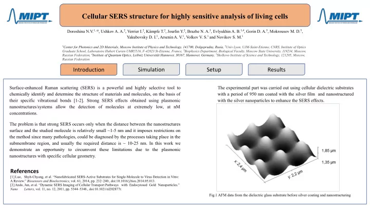
[1] Luo, Shyh-Chyang, et al. “Nanofabricated SERS-Active Substrates for Single-Molecule to Virus Detection in Vitro: A Review.” Biosensors and Bioelectronics , vol. 61, 2014, pp. 232–240., doi:10.1016/j.bios.2014.05.013. [2] Ando, Jun, et al. “Dynamic SERS Imaging of Cellular Transport Pathways with Endocytosed Gold Nanoparticles.” Nano Letters , vol. 11, no. 12, 2011, pp. 5344–5348., doi:10.1021/nl202877r. Fig.1 AFM data from the dielectric glass substrate before silver coating and nanostructuring

  2. Cellular SERS structure for highly sensitive analysis of living cells Doroshina N.V., Ushkov A. A., Verrier I., Kämpfe T., Jourlin Y., Brazhe N. A., Evlyukhin A. B., Gorin D. A., Mokrousov M. D., Yakubovsky D. I., Arsenin A. V., Volkov V. S. and Novikov S. M. Geometry of the model and mesh details Results of numerical simulations The specific geometry of the surface nanostructures and nanostructuring makes it possible to obtain considerable electromagnetic field enhancement effects in the near field, due to the normal components of the dipole moments of the particles located on the substrate (relative to the substrate, consisting of curved metal cavities). Thus it is possible to receive a strong signal from the submembrane region of bio-objects. Figure 2 shows a schematic presentation of a mitochondrion located: (A) on a flat Ag nanostructured surface with Ag nanoparticles and (B) in a cavity on Ag nanostructured surface. Results of numerical simulations are presented in Fig. 5C–F. Positions of Ag nanoparticles with diameters of 40–50 nm placed on the flat Ag surface (XY- plane) are shown in Fig. 5C. When the nanoparticle structure is irradiated by normally incident linear-polarized light beam, the electric near field above nanoparticles is very weak (Fig. 5D). When the same structure is irradiated at 65 degrees by TM-polarized light, the electric near-field above the nanoparticles increases significantly due to the presence of normal induced dipole components (Fig. 5E,F). In the plane perpendicular to the substrate surface, the near-field generated by every dipole looks like the mentioned above electric field needles (Fig. 5F). A strong contribution to the enhancement of Raman scattering is due to nanoparticles that are located on side walls of cavities of multiscale AgNSSs (Fig. 4D) which are illuminated by light of certain polarization with respect to the cavity surfaces. [3] Fig.2 Results of numerical simulations [3] Brazhe, Nadezda A., et al. “Probing Cytochrome c in Living Mitochondria with Surface-Enhanced Raman Spectroscopy.” Scientific Reports , vol. 5, no. 1, 2015, doi:10.1038/srep13793.

  3. Cellular SERS structure for highly sensitive analysis of living cells Doroshina N.V., Ushkov A. A., Verrier I., Kämpfe T., Jourlin Y., Brazhe N. A., Evlyukhin A. B., Gorin D. A., Mokrousov M. D., Yakubovsky D. I., Arsenin A. V.,Volkov V. S. and Novikov S. M. The applicability of obtained R6G was injected as a Raman active label and with nanostructures will be investigated using concentration which can not be detected by the special microcapsules with a core of ''normal' Raman. The depth of location of the R6G silicon dioxide with the inflicted layer of layer inside the microcapsule was variated by a Rhodamin 6G (R6G) on top of it and the number of polymer on top of it. It was demonstrated next layers of polymer Poly(sodium the possibility to detect SERS signal from R6G in 4-styrenesulfonate). [4] microcapsules located in cavities, while the signal from the RG6 in microcapsules located on a flat surface is not detected. The R6G was located on the depth ~10 nm in microcapsules. Fig.5 Model of the experiment [4] Mokrousov, Maksim D., et al. “Amplification of Photoacoustic Effect in Bimodal Polymer Particles by Fig. 3 Model of obtained nanostructures on the Fig.4 Model of the microcapsule with a Raman active label Self-Quenching of Indocyanine Green.” Biomedical Optics Express , vol. 10, no. 9, 2019, p. 4775., substrate doi:10.1364/boe.10.004775.

  4. Cellular SERS structure for highly sensitive analysis of living cells Doroshina N.V., Ushkov A. A., Verrier I., Kämpfe T., Jourlin Y., Brazhe N. A., Evlyukhin A. B., Gorin D. A., Mokrousov M. D., Yakubovsky D. I., Arsenin A. V.,Volkov V. S. and Novikov S. M. The cellular surfaces of the substrates were patterned via the Laser Interference Lithography [5] and covered by the Ag film (~ 100 nm) using the electron beam deposition technique. ● The next step is to apply Ag (~ 60-80 nm) nanospheres and special microcapsules to the Ag surface of the substrate. ● We expect to obtain a strong SERS effect from the special microcapsules due to the numerical simulation data and experimental theory. ● Also we planned an experiment with coatings of substrates with other plasmonic materials, such as gold and aluminum. Fig. 3 SEM image of the substrate coated with silver film doroshina.nv@phystech.edu [4] Ushkov, Andrei A., et al. “Systematic Study of Resonant Transmission Effects in Visible Band Using Variable Depth Gratings.” Scientific Reports , vol. 9, no. 1, 2019, doi:10.1038/s41598-019-51414-3.

Download Presentation
Download Policy: The content available on the website is offered to you 'AS IS' for your personal information and use only. It cannot be commercialized, licensed, or distributed on other websites without prior consent from the author. To download a presentation, simply click this link. If you encounter any difficulties during the download process, it's possible that the publisher has removed the file from their server.

Recommend


More recommend