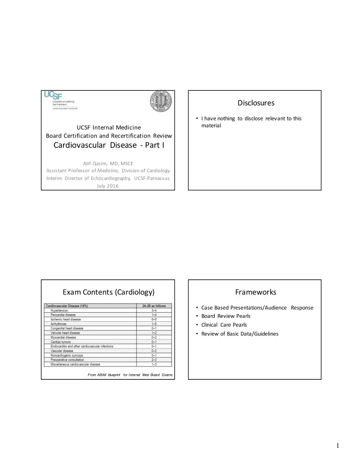
cardiovascular disease part i
play

Cardiovascular Disease - Part I Atif Qasim, MD, MSCE Assistant - PDF document

Disclosures I have nothing to disclose relevant to this material UCSF Internal Medicine Board Certification and Recertification Review Cardiovascular Disease - Part I Atif Qasim, MD, MSCE Assistant Professor of Medicine, Division of


  1. Disclosures • I have nothing to disclose relevant to this material UCSF Internal Medicine Board Certification and Recertification Review Cardiovascular Disease - Part I Atif Qasim, MD, MSCE Assistant Professor of Medicine, Division of Cardiology Interim Director of Echocardiography, UCSF-Parnassus July 2016 Exam Contents (Cardiology) Frameworks • Case Based Presentations/Audience Response • Board Review Pearls • Clinical Care Pearls • Review of Basic Data/Guidelines From ABIM blueprint for Internal Med Board Exams 1

  2. Cardiac Exam Pearls Modern Medicine and the Heart 1. Blood pressure: – Severe Hypertension (e.g. 220/110) à think aortic dissection • Chest pain? Short of breath? – Large pulse pressure with low diastolic (e.g. 180/50) à think aortic regurgitation – EKG 2. Carotids: – Labs (BNP, troponin) – Weak and slow (a.k.a. parvus et tardus ) à think aortic stenosis – Bifid (double peak) pulse à think HOCM – Chest X-Ray 3. JVP: – CT scan – Large V wave à tricuspid regurgitation – Echo – Rapid Y descent à constriction 4. PMI – Stress T esting - Laterally displaced à a dilated heart, systolic heart failure – Cardiac Cath - Sustained, non displaced à a thick heart, diastolic heart failure Cardiac Exam Pearls (2) Cardiac Exam Pearls (3) 1. First heart sound ( closure of mitral and tricuspid valves ) : Murmurs: Soft S1 à think mitral (or tricuspid) regurgitation – Location, location, location A P Loud S1 à think mitral stenosis – Location and timing in cardiac T 2. Second heart sound (closure of aortic and pulmonic valves) : M cycle will tell you the cause Physiologically split i.e. split only during inspiration à norma l – Fixed splitting à think ASD – Holosystolic – MR, TR, VSD Loud P2 à think pulmonary hypertension – – Soft A2 (or Single S2) à Severe Aortic stenosis. Ejection – AS, PS, HCM Mid to late systolic –MVP with MR 3. S3 à think volume overload, dilated heart Rare* non-valvular murmurs: 4. S4 à think pressure overload, thick heart Continuous “machinery” think patent ductus arteriosus (PDA) 2

  3. Dynamic Maneuvers HOCM and Valsalva Maneuver Effect Reason Inspiration Augments right-sided Due to increased venous murmurs filling of right heart Valsalva AugmentsHOCM murmur . Increases intrathoracic Makes MVP (mitral valve pressure, compresses LV prolapse) click earlier and reducing LV volume. longer associated MR Reduces preload and murmur . LVEDP Squatting MakesHOCM murmur Increases arterial softer, MVP click later, all resistance, venous return other murmurs louder . (preload) and stroke volume and chamber size Rapid standing Opposite of squatting Opposite of squatting Image courtesy of www.studyblue.c om Cardiac Examination Pearls (4) MVP and Valsalva • There are often clues in the history!!!! – Young patients à think congenital heart disease – Old patients à think degenerative valve disease – Foreign patients à think rheumatic heart disease – Post-MI à think mechanical complications (VSD or mitral regurgitation) – Cancer patients à think about pericardial Valsalva disease, chemo related cardiomyopathy From O'Rourke RA, Crawford MH. The systolic click-murmur syndrome: clinical recognition and management. Curr Probl Cardiol 1976;1:1–60 . 3

  4. Approach to Tachycardia Approach to ECGs for the boards • Rate, rhythm, axis Irregular = AFIB Narrow = • P-Q-R-S-T conducts via normal Regular or pathways (i.e. irregular? Check P-waves: • P= atrial depolarization supraventricular Regular = 1. normal = in origin) Sinus Sinus tach , QRS • PR = AV conduction tach, A 2. sawtooth = narrow Flutter or Aflutter , or wide SVT 3. none or • QRS = ventricular Wide = abnormal = SVT ventricular origin VT or SVT depolarization or conducts via with abnormal aberrancy • T = ventricular pathway repolarization Approach to Bradycardia Approach to Cases Really only 3 things it is likely to be on this exam: • Read the question first • Sinus bradycardia • Heart block (AV nodal block) – I will present this way • Slow Afib • Figure out what question type So ask yourself: – which diagnostic test 1. Are there P waves? – management decision – If no, it is probably Afib 2. Is every P wave followed by a QRS complex? – you make the diagnosis – If yes, sinus brady • Read vignette with question in mind, highlight st or 2 nd degree heart block If no, complete heart block >> 1 – any clues. Exam pearl: Examiners like to stop meds – may need to stop beta-blockers 4

  5. Case 1: 22 year old man with Case 1: What is the next best test? exertional syncope A. T readmill Exercise ECG Stress Presented to ER after an episode of sudden loss of consciousness immediately after a 3 mile jog. He B. Cardiac Event monitor awoke completely after 3-4 minutes. Denied chest pain, shortness of breath or palpitations. C. Dobutamine stress echocardiography • FH: positive for sudden death in a cousin D. T ransthoracic echocardiogram E. Serial troponin I levels and EKGs • PMH: Uncomplicated rotator cuff repair • PE: HR =60, BP = 90/60, JVP = 6 cm, S4 gallop, no murmur. Lungs clear. No edema. Case 1: EKG Case 1: What is the next best test? A. T readmill Exercise ECG Stress B. Cardiac Event monitor C. Dobutamine stress echocardiography D. T ransthoracic echocardiogram E. Serial troponin I levels and EKGs 5

  6. Case 1: EKG Review Case 1: What is the next best test? A. T readmill Exercise ECG Stress B. Cardiac Event monitor C. Dobutamine stress echocardiography D. Transthoracic echocardiogram E. Serial troponin I levels and EKGs Case 1: Educational Objectives Overall Causes of Syncope – Know the causes and recommended evaluation • Cardiac: 18% for syncope • Neurologic: 10% • Vasovagal: 24% – Recognize the symptoms and signs of hypertrophic cardiomyopathy • Orthostatic: 8% • Medications: 3% – Know the risk factors for sudden death in a patie nt • Unknown: 37% with hypertrophic cardiomyopathy 6

  7. Cardiac Syncope Syncope Evaluation • Structural causes • Arrhythmic causes • Careful history and physical examination crucial in work-up of syncope – Common – Bradycardia – Reveals probable cause in 50% of patients • AS - usually exertional • Sinus bradycardia • Hypertrophic obstructive – Vasovagalsyncope is strongly suggested by characteristic • AV block cardiomyopathy - often prodrome and may be confirmed by Tilt table test • Carotid sinus post-exercise or due to hypersensitivity • EKG arrhythmias – Tachycardia – Less common • Echo indicated with h/o heart disease, abnormal exam or • SVT (rare) abnormal EKG or elderly • PE, dissection, tamponade • VT – Uncommon • Holter or event monitor • Tilt Table • Pulmonary HTN, atrial myxoma Exertional syncope often ominous Hypertrophic Cardiomyopathy(HCM) • Differential diagnosis: • Autosomal dominant inheritance pattern with – Obstruction to left ventricular outflow (aortic stenosis an d variable penetrance. 1/500 incidence. hypertrophic cardiomyopathy [HOCM]) • Usually presents in adolescent or young adult – Ventricular tachycardia but can present at any age – Pulmonary hypertension • Triad of symptoms: – Neurocardiogenic syncope • T readmill Exercise ECG Stress T esting should be Chest pain, Dyspnea and Syncope delayed until structural causes excluded 7

  8. HCM: Physical Examination HCM (cont’d) • EKG: L VH with repolarization abnormalities • Non-obstructive: S4, sustained apical impulse • Echocardiogram: • Classic: Left ventricular hypertrophy with septal >>posterior wa ll • Obstructive: thickness (asymmetric septal hypertrophy) • Obstruction: associated with systolic anterior motion (SAM) of • Bisferiens carotid pulse (“spike and dome”) the mitral valve. Sometimes there is associated MR. • Double or Triple apical impulse • Pharmacologic treatment • Systolic murmur that increases with standing or strain phase of • Beta-blocker Valsalva and decreases with passive leg raising or squatting, • Calcium channel blocker (Verapamil, Diltiazem) increases post-PVC • Avoid any vasodilator or positive inotropic agent (Dobutamine • Dynamic obstruction: echocardiography relatively contraindicated) • Occasionally may hear murmur only with maneuver • Avoid dehydration • See obstruction only with exercise HOCM and systolic anterior motion HCM: Other Treatment • Avoid high intensity sports • Surgical myomectomy or alcohol septal ablation in obstructive form only in pts refractory to med Rx • ICD for those at high risk for sudden death if : • Prior cardiac arrest • One or more family members with sudden death caused by HCM, • Syncope otherwise unexplained, especially if related to physical activity, occurs repeatedly, or appears in young people. • Ventricular tachycardia on Holter monitor • Blood pressure fails to rise during exercise testing, esp if younger than 50 years. • Extreme thickness of LV septum (30 mm or more) on echo. 8

Download Presentation
Download Policy: The content available on the website is offered to you 'AS IS' for your personal information and use only. It cannot be commercialized, licensed, or distributed on other websites without prior consent from the author. To download a presentation, simply click this link. If you encounter any difficulties during the download process, it's possible that the publisher has removed the file from their server.

Recommend


More recommend