
SLIDE 1
5/23/2014 1
Beyond Acute Appendicitis:
Fascinating Lesions of the Vermiform Appendix
Laura W. Lamps, M.D. University of Arkansas for Medical Sciences Little Rock, AR
The Appendix: historical perspectives
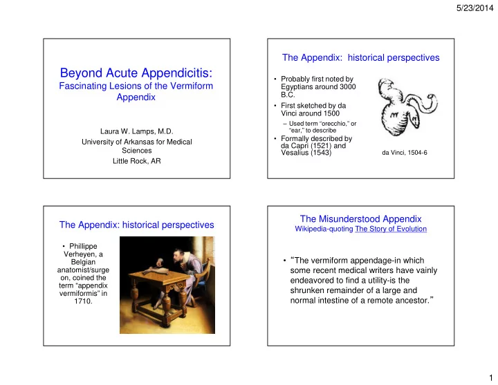
- Probably first noted by
Egyptians around 3000 B.C.
- First sketched by da
Vinci around 1500
– Used term “orecchio,” or “ear,” to describe
- Formally described by
da Capri (1521) and Vesalius (1543)
da Vinci, 1504-6
The Appendix: historical perspectives
- Phillippe
Verheyen, a Belgian anatomist/surge
- n, coined the
term “appendix vermiformis” in 1710.
The Misunderstood Appendix
Wikipedia-quoting The Story of Evolution
- “The vermiform appendage-in which