
SLIDE 1
10/12/2018 1
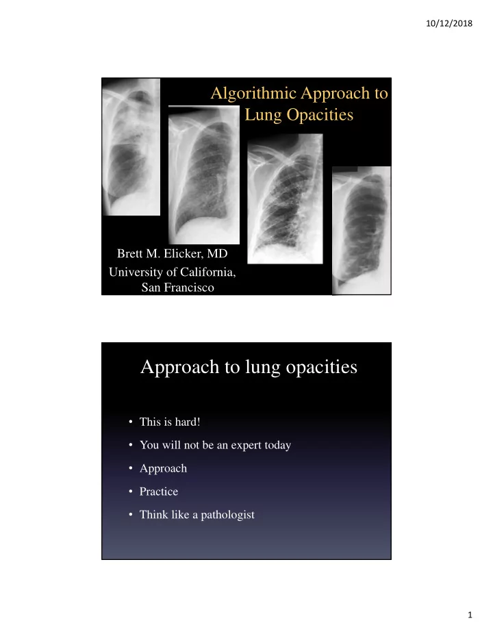
Algorithmic Approach to Lung Opacities
Brett M. Elicker, MD University of California, San Francisco
Approach to lung opacities
- This is hard!
- You will not be an expert today
- Approach
- Practice
- Think like a pathologist