
SLIDE 1
Proceedings of UCLA Healthcare
- VOLUME 18 (2014)-
CLINICAL VIGNETTE
An Unusual Presentation of Scrotal Calcinosis: Painful on Exercise and Originating from a Hybrid Cyst
Nima M. Gharavi, MD, PhD1, Jennifer T. Hau, MD2*, and David P. Beynet, MD2
1Cedars-Sinai Medical Group, Department of Dermatology, Los Angeles, California, USA 2Division of Dermatology, David Geffen School of Medicine, University of California, Los Angeles, California, USA
*Correspondence to: Jennifer T. Hau, MD University of California, Los Angeles, Division of Dermatology 200 Medical Plaza, Suite 465 Los Angeles, CA 90095-6957 P: (310) 825-6911 F: (310) 794-7005 E: jhau@mednet.ucla.edu Introduction Scrotal calcinosis is an uncommon, benign disorder characterized by the progressive development of calcified nodules within the dermis of the scrotal skin. The condition typically affects adolescent to middle-aged males without abnormalities in calcium or phosphorous metabolism. Nodules may be solitary or multiple and range from 1 millimeter to several centimeters in size. The lesions are usually firm and asymptomatic, but associated symptoms including pruritus and pain have been reported. There is no consensus on the etiology of scrotal calcinosis. The condition is considered idiopathic by some authors. Other suggested pathogenic mechanisms include the dystrophic calcification of preexisting epidermal or eccrine cysts. Case report A 32-year-old Caucasian man presented with multiple nodules
- n the scrotum, which had progressively increased in size and
number over a period of several years. The nodules were painful with exercise, and the pain resolved with rest. The patient had no history of trauma, sexually transmitted infection, drug abuse, or any inflammatory, endocrinologic, neoplastic, or metabolic disorders. His family history was unremarkable. Physical examination revealed twenty firm, tender, yellow- white, well-circumscribed, 2 to 5 mm nodules present on the
- scrotum. The associated pain and the cosmetic appearance of
the nodules prompted the patient to request removal of the
- lesions. All of the lesions were surgically excised in a day-
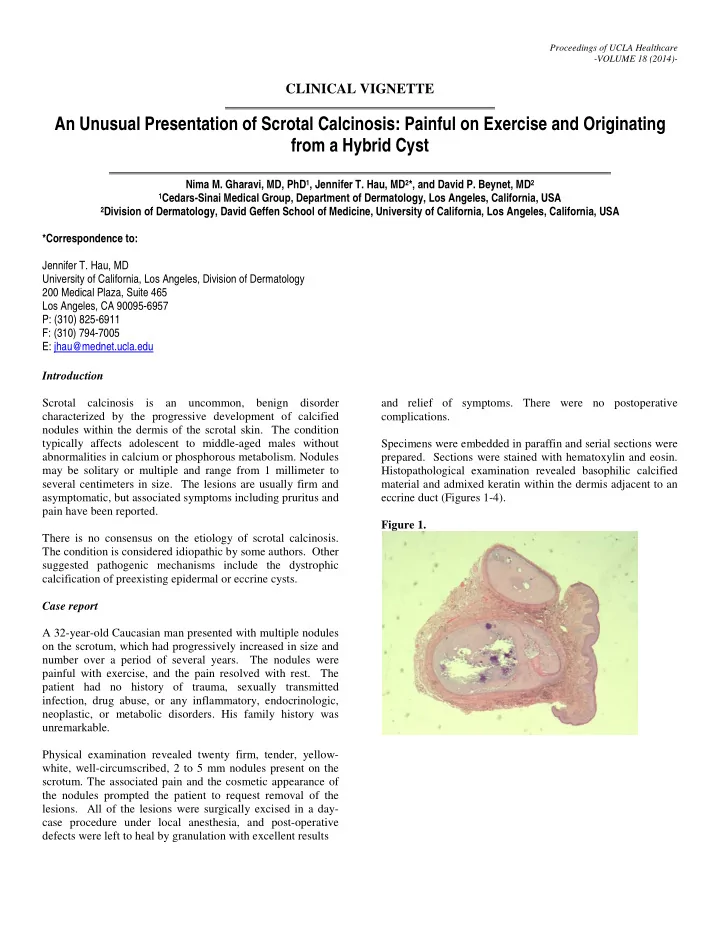
case procedure under local anesthesia, and post-operative defects were left to heal by granulation with excellent results and relief of symptoms. There were no postoperative complications. Specimens were embedded in paraffin and serial sections were
- prepared. Sections were stained with hematoxylin and eosin.