An Unusual Presentation of luxation of the globe, forced globe - - PDF document

an unusual presentation of
SMART_READER_LITE
LIVE PREVIEW

An Unusual Presentation of luxation of the globe, forced globe - - PDF document

Alim S, et al., J Ophthalmic Clin Res 2016, 3: 020 DOI: 10.24966/OCR-8887/100020 HSOA Journal of Ophthalmology and Clinical Research Case Report An Unusual Presentation of luxation of the globe, forced globe rotation or sudden rise in


slide-1
SLIDE 1

Introduction

Optic nerve avulsion is very rare condition and it is an important complication of blunt trauma to the globe. Tie optic nerve avulsion is most frequently secondary to motor vehicles and bicycle accidents followed by falls [1]. Sport injuries such basketball, cricket and skateboarding are rarely caused optic nerve avulsion [2]. Complete avulsion of the optic nerve mechanisms defjned such as anterior

*Corresponding author: Sait Alim, Department of Ophthalmology, Gaziosman- paşa University, Tokat, Turkey, Tel: +90 5073969257; Fax: +90 3562521625; E-mail: drsaitalim@gmail.com Citation: Alim S, Demir S, Gökçe E, Ortak H, Demir HD, et al. (2016) An Unusual Presentation of Optic Disc Drusen with Optic Nerve Avulsion. J Ophthalmic Clin Res 3: 020. Received: January 29, 2016; Accepted: March 30, 2016; Published: April 13, 2016

luxation of the globe, forced globe rotation or sudden rise in intraocular pressure [3]. Optic disc drusen or optic disc head drusen are found about 2.4% in a necropsy study [4]. About two thirds to three quarters of clinical cases are bilateral. Here we present a patient with bilateral optic disc with drusen who had a blunt trauma to his lefu eye by a crane lifuing chain which caused complete optic nerve avulsion.

Case Report

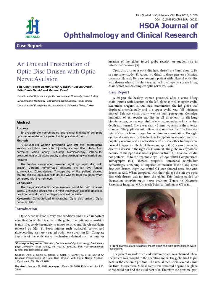
A 50-year-old healthy woman presented afuer a crane lifuing chain trauma with luxation of the lefu globe as well as upper eyelid lacerations (Figure 1). On local examination the lefu globe was displaced anterolaterally and the upper eyelid was full thickness

  • incised. Lefu eye visual acuity was no light perception. Complete

limitation of extraocular motility in all directions. In slit-lamp biomicroscopy, cornea was minimal edematous and anterior chamber depth was normal. Tiere was nearly 3 mm hyphema in the anterior

  • chamber. Tie pupil was mid-dilated and non-reactive. Tie Lens was
  • intact. Vitreous hemorrhage obscured fundus examination. Tie right

eye visual acuity was 10/10 in Snellen. Except for an absent consensual pupillary reaction and an optic disc with drusen, other fjndings were normal (Figure 2). Ocular Ultrasonography (US) showed an optic disc with drusen in the right eye (Figure 3). Tie globe was hypotonic because of the optic disc head separation from it. Tierefore we did not perform US to the hypotonic eye. Lefu eye orbital Computerized Tomography (CT) showed proptosis, intraconal retrobulbar hemorrhage, stretching of superior extraocular muscle and optic disc with drusen. Right eye orbital CT scan showed optic disc with drusen as well. When compared with the right eye the lefu eye optic disc with drusen was far from the globe. Tiis fjnding guided us diagnosing complete optic nerve avulsion (Figure 4). Magnetic Resonance Imaging (MRI) revealed similar fjndings as CT scan. Tie patient was informed and written consent was obtained. Tien, the patient was brought to the operating room. Tie globe tried to put back in the anatomic position. Tie medial rectus was severed 2 mm far from its insertion. Medial rectus was retracted beyond the globe so we could not fjnd the distal part of it. Tierefore the proximal part

Alim S, et al., J Ophthalmic Clin Res 2016, 3: 020 DOI: 10.24966/OCR-8887/100020

HSOA Journal of Ophthalmology and Clinical Research

Case Report

Abstract

Purpose To evaluate the neuroimaging and clinical fjndings of complete

  • ptic nerve avulsion of a patient with optic disc drusen.

Methods A 50-year-old woman presented with left eye anterolateral luxation and vision loss after injury by a crane lifting chain. Best corrected vision acuity, slit-lamp biomicroscopy, intraocular pressure, ocular ultrasonography and neuroimaging was carried out. Results The fundus examination revealed right eye optic disc with

  • drusen. Vitreous hemorrhage obscured the left eye fundus
  • examination. Computerized Tomography of the patient showed

that the left eye optic disc with drusen was far from the globe when compared with the right eye. Conclusion The diagnosis of optic nerve avulsion could be hard in some

  • cases. Clinicians should keep in mind that in such cases if optic disc

head contains drusen the diagnosis would be easier. Keywords: Computerized tomography; Optic disc drusen; Optic nerve avulsion Sait Alim1*, Selim Demir1, Erkan Gökçe2, Hüseyin Ortak1, Helin Deniz Demir1 and Mehmet Esen3

1Department of Ophthalmology, Gaziosmanpaşa University, Tokat, Turkey 2Department of Radiology, Gaziosmanpaşa University, Tokat, Turkey 3Department of Emergency, Gaziosmanpaşa University, Tokat, Turkey

An Unusual Presentation of Optic Disc Drusen with Optic Nerve Avulsion

Figure 1: Anterolateral luxation of the left globe and full thickness upper eyelid lacerations.

slide-2
SLIDE 2

Citation: Alim S, Demir S, Gökçe E, Ortak H, Demir HD, et al. (2016) An Unusual Presentation of Optic Disc Drusen with Optic Nerve Avulsion. J Ophthalmic Clin Res 3: 020.

  • Page 2 of 3 •

J Ophthalmic Clin Res ISSN: 2378-8887, Open Access Journal DOI: 10.24966/OCR-8887/100020

Volume 3 • Issue 1 • 100020

  • f the medial rectus sutured to the bone periosteum of medial wall
  • f the globe. Tie remainder of other rectus muscles insertions was
  • intact. But the superior rectus was stretched and so it folded over itself

and sutured. In the end the upper eyelid was repaired. Postoperative- ly the globe was in anatomic position and the patient’s visual acuity was no light perception. Eye movements were restricted. Tie right eye examination was normal. It is a very rare case and to the best of our knowledge it is the fjrst case evaluating optic disc head drusen with

  • ptic nerve avulsion.

Discussion

In optic nerve avulsion, the optic nerve is forcibly detached from the globe and usually there is an object that intrudes between the

  • rbit wall and globe and dislocates the eye. It is thought that, sudden

severe rotation of the globe, anterior displacement of the eyeball and rapid rise in intraocular pressure leading to expulsion of optic nerve

  • ut of scleral canal can cause optic nerve avulsion [3]. Tie luxation
  • f the globe is seen rarely and following trauma medial rectus is the

most frequently ruptured extraocular muscle [5,6]. Our patient had a trauma from medial wall of the globe and the crane lifuing chain displaced the globe anteriorly. Tie eye was luxated and the medial rectus was severed near its insertion. If there is a clear media the diagnosis of complete optic nerve avulsion can be done easily. In such cases the disc area will be seen empty. If there is a vitreous hemorrhage

  • r mature cataract then it would be diffjcult to diagnose it. In such

cases it is essential to diagnosis the patient properly to avoid giving inappropriate treatment such as high-dose steroids or optic nerve decompression [3]. Optic nerve drusen is defined as hyaline bodies that result from calcification of intracellular axonal mitochondria located in the prelaminar portion of optic nerve [4]. B-scan US, a non-invasive diagnostic tool, is a reliable method for confirming the presence of

  • ptic disc drusen [7]. CT is also a useful method for diagnosis of optic

disc drusen due to their calcium content. Various diagnostic tests like MRI, CT scan, US and fundus fmuorescein angiography have been used in diagnosis of optic nerve

  • avulsion. Tiese diagnostic tests could not always show the optic

nerve avulsions. For this reason sometimes surgical exploration may needed for exact diagnosis of optic nerve avulsion. Roth et al., [8] have described a patient with optic nerve avulsion who had trauma to his eyeball by the golf club. Tie CT scan of brain and orbits showed no abnormalities. Foster et al., [9] carried out a study to fjnd out the role of diagnostic imaging in patients with optic nerve avulsion. Neuroimaging including CT scan and MRI or both was done for six patients. Only in one case disinsertion of optic nerve from the globe was discovered. In two cases MRI could not reveal optic nerve

  • avulsion. Complete optic nerve avulsion was seen just in one case

when the globe was surgically explored. Tierefore, it is usually hard to diagnosis optic nerve avulsion by CT scan and MRI. Our patient was also asked for orbital CT scan and MRI. Because of retrobulbar hemorrhage MRI fjndings was not helpful. Orbital CT scan revealed

  • ptic disc with drusen. While compared with the right eye the lefu eye
  • ptic disc with drusen was far from the globe (Figure 3). Tiis fjnding

leaded us to the diagnosis of complete optic nerve avulsion. US is also may be useful in diagnose of optic nerve avulsion. Prior studies of US have revealed encouraging results in diagnosis of optic nerve avulsion [10]. Sawhney et al., [3] had found that US of globe demonstrated hypoechoic defect in the posterior ocular coat showing complete avulsion in their two cases.

Conclusion

Optic nerve avulsion is sight threatening complication of blunt

  • trauma. It is very important to expose optic nerve avulsion because

treatment should be done according to the diagnosis. Tie purpose of the present study was to emphasize the importance of optic disc with drusen in diagnosis of complete optic nerve avulsion.

Figure 2: Fundus photograph of the right eye with optic disc drusen. Figure 3: Ultrasonograpy of the right eye. Optic disc drusen showed by white arrow. Figure 4: Computerized tomography scan of the patient. Bilateral optic disc drusen showed by black arrow. As it is seen in the CT scan the drusen is far from the left globe. Posterior border of the left globe is shown by white arrow.

slide-3
SLIDE 3

Citation: Alim S, Demir S, Gökçe E, Ortak H, Demir HD, et al. (2016) An Unusual Presentation of Optic Disc Drusen with Optic Nerve Avulsion. J Ophthalmic Clin Res 3: 020.

  • Page 3 of 3 •

J Ophthalmic Clin Res ISSN: 2378-8887, Open Access Journal DOI: 10.24966/OCR-8887/100020

Volume 3 • Issue 1 • 100020

References

  • 1. Steinsapir KD, Goldberg RA (1994) Traumatic optic neuropathy. Surv Oph-

thalmol 38: 487-518.

  • 2. Roberts SP, Schaumberg DA,Thompson P (1992) Traumatic avulsion of the
  • ptic nerve. Optom Vis Sci 69: 721-727.
  • 3. Sawhney R, Kochhar S, Gupta R, Jain R, Sood S (2003) Traumatic optic

nerve avulsion: role of ultrasonography. Eye (Lond) 17: 667-670.

  • 4. Friedman AH, Henkind P, Gartner S (1975) Drusen of the optic disc. A histo-

pathological study. Trans Ophthalmol Soc UK 95: 4-9.

  • 5. Ersan I, Adam M, Oltulu R, Zengin N, Okka M (2016) Traumatic luxation of

the globe: A 6-year follow-up. Orbit 35: 69-71.

  • 6. O’Toole L, Long V, Power W, O’Connor M (2004) Traumatic rupture of the

lateral rectus. Eye (Lond) 18: 221-222.

  • 7. Zaouali S, Abroug N, Khochtali S, Kahloun R, Jelliti B, et al. (2014) Optic

nerve head drusen: a comparative study of 10 MHz and 20 MHz ultrasound

  • probes. Int Ophthalmol 35: 229-232.
  • 8. Roth DB, Warman R (1999) Optic nerve avulsion from a golfjng injury. Am J

Ophthalmol 128: 657-658.

  • 9. Foster BS, March GA, Lucarelli MJ, Samiy N, Lessell S (1997) Optic nerve
  • avulsion. Arch Ophthalmol 115: 623-630.
  • 10. Talwar D, Kumar A, Verma L, Tewari HK, Khosla PK (1991) Ultrasonography

in optic nerve head avulsion. Acta Ophthalmol (Copenh) 69: 121-123.