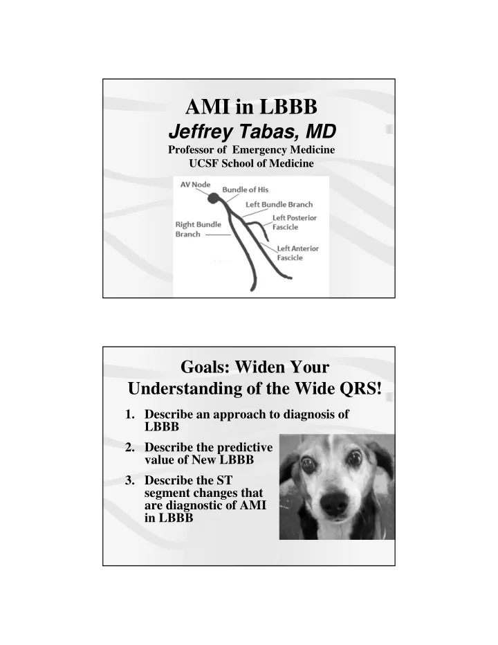
ami in lbbb
play

AMI in LBBB Jeffrey Tabas, MD Professor of Emergency Medicine - PDF document

AMI in LBBB Jeffrey Tabas, MD Professor of Emergency Medicine UCSF School of Medicine Goals: Widen Your Understanding of the Wide QRS! 1. Describe an approach to diagnosis of LBBB 2. Describe the predictive value of New LBBB 3. Describe


  1. AMI in LBBB Jeffrey Tabas, MD Professor of Emergency Medicine UCSF School of Medicine Goals: Widen Your Understanding of the Wide QRS! 1. Describe an approach to diagnosis of LBBB 2. Describe the predictive value of New LBBB 3. Describe the ST segment changes that are diagnostic of AMI in LBBB

  2. Case 1 65 y.o F with fatigue • 65 y.o. F with the sugar diabetes BIBA w/ fatigue and vomiting for a few hours. • Vital signs and physical exam are unremarkable 1) 65 y.o. F with fatigue

  3. Case 1 65 y.o F with fatigue • No Old ECG available • Called for records to another hospital and faxed consent • While awaiting response, patient went into Vfib, was resuscitated, rushed to cath and found to have 100% LAD Case 1 65 y.o F with fatigue 3 Questions 1. Is this LBBB? 2. Is this NEW LBBB? 3. Can we read ST segment abnormalities?

  4. 1) Is this LBBB? 6 CAUSES - WIDE QRS  Bundle branch block  Ventricular rhythm  Hyperkalemia  Medications  Paced rhythm  WPW 1) Is this LBBB?  The QRS is wide, usually > 0.14  Look at TERMINAL portions of the QRS in Lead V1 and Lead 1 (V6)  LBBB = Terminal R in 1 (V6) and Slurred S in V1

  5. Left Bundle Branch Man LBBB Man  Left hand is up for LBBB  Left hand represents left side - lateral leads  Right hand represents right side – V1  Hand points in direction of the final wave of the QRS (i.e. R wave points up, Q and S waves point down

  6. LBBB 2) Is this NEW LBBB? Indications for PCI and Thrombolytics • 1mm ST elevation in 2 contiguous leads or • Left Bundle Branch not known to be old

  7. Predictive Value of New or Presumed New LBBB Chang, Am JEM, 2009 • 55 with New LBBB = 7.3% AMI • 136 with Old LBBB = 5.2% AMI • 7746 with no LBBB = 6.1% AMI New LBBB is not predictive of AMI 2) Is this NEW LBBB? Indications for PCI and Thrombolytics • 1mm ST elevation in 2 contiguous leads or 2013 ACCF/AHA Guideline for the Management • Left Bundle Branch not known to be old of ST-Elevation Myocardial Infarction “New or presumably new LBBB at presentation occurs infrequently, may interfere with ST-elevation analysis, and should not be considered diagnostic of acute MI in isolation.”

  8. 3) Can we read the ST segments (i.e. Dx AMI) in LBBB? 2013 ACCF/AHA Guideline for the Management of ST-Elevation Myocardial Infarction • Criteria for ECG diagnosis of acute STEMI in the setting of LBBB have been proposed (see Online Data Supplement 1 ) LBBB: Normal ST Segments  Iso-electric or  Discordant (ST segment opposite the terminal QRS)  This is true for every lead

  9. LBBB ACUTE MI in LBBB CONCORDANT CONCORDANT ST Elevation ST Depression

  10. ACUTE MI in LBBB EXCESSIVE DISCONCORDANCE ST:S wave = 0.25 or more Acute MI in LBBB Annals of EM, October 2008

  11. Acute MI in LBBB • 1 mm Concordant ST elevation – 10 studies with 1,614 patients – Sensitivity = 20% (NLR = 0.8) – Specificity of 98% (PLR = 7.9) • 5 mm Discordant ST elevation – Specificity of 80% (PLR = 4.5) Acute MI in LBBB Annals of EM, August 2012

  12. ST segments in AMI/LBBB • Excessive Discordance – ST elevation: S wave >= 1:4 – ST depression: R wave >= 1:4 – Significant improvement in sensitivity and specificity 1) 65 y.o. F with fatigue

  13. 1) 65 y.o F with fatigue – baseline LBBB Another pt with LBBB and Chest Pain c

  14. Yet another pt with LBBB and Chest Pain c ACUTE MI in Paced Rhythms  Same as with LBBB!

  15. 80 y.o. M with CP and pacer Prior ECG

  16. Take Home Points Dx of AMI in LBBB 1. Determine if LBBB – LBBB man 2. Do not use New LBBB to predict AMI Take Home Points Dx of AMI in LBBB 3. Determine if AMI is present Expected ST segments – Opposite terminal R or S wave – or isoelectric – in every lead

  17. Take Home Points Dx of AMI in LBBB 3. Determine if AMI is present Acute MI 1 mm Concordant ST segments (in same • direction as last wave of QRS) in any lead Excessive Discordance of ST segments • (opposite to terminal R or S wave) – ST:S wave ratio > = 1:4 Treatment of Chest Pain with LBBB or a Paced Rhythm • If ST changes diagnostic of AMI then – Reperfuse immediately (Lytics or Cath Lab) if • If no concerning ST changes then – Involve cardiology consultant early if possible – Reperfuse for high suspicion of STEMI (> 50%?) – Use cardiac markers or formal echo to rule out AMI in the rest

Download Presentation
Download Policy: The content available on the website is offered to you 'AS IS' for your personal information and use only. It cannot be commercialized, licensed, or distributed on other websites without prior consent from the author. To download a presentation, simply click this link. If you encounter any difficulties during the download process, it's possible that the publisher has removed the file from their server.

Recommend


More recommend