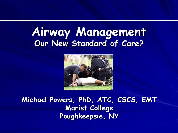
SLIDE 1
Airway Management
Our New Standard of Care?
Michael Powers, PhD, ATC, CSCS, EMT Marist College Poughkeepsie, NY
SLIDE 2
SLIDE 3
SLIDE 4
“Fill your hands you son of a bitch!”
SLIDE 5 From all accounts, Adam Seymour was in shape and impressing his coaches. But on the last lap
- f a mile and a half run, Seymour collapsed.
Trainer was one of the first people to respond, giving Seymour CPR until rescue crews arrived. "We are trained to do this type of activity but you never picture in your head that you are actually going to use those skills," said .
SLIDE 6
Are you fully prepared?
SLIDE 7
Risk Management
Wheeler's mother to seek 'substantial' damages EVANSTON, Ill. -- The mother of Northwestern football player Rashidi Wheeler plans to file a lawsuit against the university later this week seeking "substantial" damages over his death in a preseason conditioning drill.
SLIDE 8
Are you fully prepared?
SLIDE 9
Airway Management
Accessing the airway
– Equipment issues Helmets Facemasks
SLIDE 10
Airway Management
Opening the airway
– Non-trauma: head-tilt-chin-lift – Trauma: jaw thrust or modified jaw thrust – A patient without an airway is a dead patient
SLIDE 11
Airway Management
Airway Adjuncts
– Oropharyngeal Airway (OPA) Assist in maintaining an open airway on unresponsive patients without a gag reflex Patients with a gag reflex will vomit Size: measure from the corner of the lips to the bottom of the earlobe or angle of mandible
SLIDE 12
Airway Management
Airway Adjuncts
– Oropharyngeal Airway Insert airway with the tip facing toward the roof of the patient's mouth Advance the airway gently until resistance is encountered then turn it 180° so that it comes to rest with the flange on the patient's teeth
SLIDE 13
Airway Management
Airway Adjuncts
– Oropharyngeal Airway Contraindications Patient is conscious. Patient has a gag reflex. There is some foreign body that is blocking the airway, such as food, dentures, etc that should be removed first if possible.
SLIDE 14 Airway Management
Airway Adjuncts
– Nasopharyngeal Airway (NPA) Used when patient’s have an intact gag reflex An NPA is less likely to stimulate vomiting than an oral airway and may also be used on patients who are responsive, but still need assistance in keeping the tongue from
SLIDE 15
Airway Management
Airway Adjuncts
– Nasopharyngeal Airway (NPA) The nasal airway is a pliable tube that is inserted through the nose that when fully inserted, the tip is located in the posterior pharynx To appropriately select the size, measure from the tip of the nose to the tip of the patient’s ear One thing to keep in mind is the diameter of the airway in relation to the patient’s nostril.
SLIDE 16
Airway Management
Airway Adjuncts
– Nasopharyngeal Airway (NPA) Once the appropriately sized device has been chosen, lubricate the airway with a water soluble lubricant The airway will then be inserted into the nostril with the bevel pointed toward the septum
SLIDE 17
Airway Management
Airway Adjuncts
– Nasopharyngeal Airway (NPA) Gently insert the device until the flange is resting atop the patient’s nostril If the airway does not insert fully into the nostril then attempt the same procedure in the opposite nostril
SLIDE 18
Airway Management
Airway Adjuncts
– Nasopharyngeal Airway Contraindications Patient with significant head trauma. Patient with nasal fractures. (inserting a NPA in a patient with either of these problems could result with the NPA being inserted into the brain)
SLIDE 19 Airway Management
Suctioning
Be sure to use the rigid catheter for mouths and a bulb suction or French catheter for nasal passages
- 2. Insert the catheter into the oral cavity
without suction (begin at the base of the tongue and work anterior)
SLIDE 20 Airway Management
Suctioning
- 3. Apply suction for no more than 15 seconds
For children, try to shorten suction time When the pt. has emesis, sputum, or saliva, that cannot be removed quickly, the pt. should be log rolled and the oropharynx should be cleared manually
- 4. Artificially ventilate, and then suction for
another 15 seconds. Continue this operation as needed.
SLIDE 21
Airway Management
Artificial Ventilation
– Mouth to mask (can have O2 connected) – Bag valve mask (BVM)
SLIDE 22
Airway Management
Oxygen Delivery
– Non-rebreather mask – Nasal cannula
SLIDE 23
Airway Management
Supralaryngeal airways
– Combitube – King – Laryngeal mask airway (LMA)
SLIDE 24
Airway Management
Esophageal-Tracheal Combitube™ (dual- lumen tube)
– Patient is unconscious and no apparent gag reflex – Adult combitube - for patients above 5' tall (41 French) – Small adult combitube - for patients between 4' and 6' tall (37 French or combitube SA)
SLIDE 25
Airway Management
Combitube contraindications
– Responsive patients with an intact gag reflex – Patients with known esophageal disease – Patients who have ingested caustic substances – Known or suspected foreign body obstruction of the larynx or trachea – Presence of a tracheotomy
SLIDE 26
Airway Management
Combitube insertion
– Patent airway and ventilation should already have been established by other basic methods – Hyperventilate – Lubricate the tube – Insert the thumb of a gloved hand into the patient's mouth, grasping the tongue and mandible between the thumb and index finger, and lift upward
SLIDE 27
Airway Management
Combitube insertion
– With the other hand, hold the Combitube with the curve in the same direction as the curve of the pharynx and insert the tip into the mouth – Advance carefully until the printed ring is aligned with the teeth
SLIDE 28
Airway Management
Combitube insertion
– DO NOT FORCE THE COMBITUBE!. If the tube does not advance easily, redirect it or withdraw and reinsert – If the Combitube is not successfully placed within 30 seconds, remove the device and hyperventilate the patient for 30 seconds before re-attempting insertion
SLIDE 29
Airway Management
Combitube insertion
– Inflate line 1, blue pilot balloon leading the pharyngeal cuff, with 100ml of air using the 140ml (cc) syringe. (This may cause the Combitube to move slightly from the patient's mouth) – Inflate line 2, white pilot balloon leading to the distal cuff, with approximately 15ml of air using the 20ml (cc) syringe
SLIDE 30
Airway Management
Combitube insertion
– Begin ventilation through the longer blue (distal) tube and watch for chest rise – If auscultation of breath sounds is positive and auscultation of gastric air sounds is negative, continue ventilation. – If no chest rise, negative lung sounds, and/or positive gastric air sounds with ventilation through the distal tube, begin ventilation through the shorter clear (proximal) tube.
SLIDE 31
Airway Management
Combitube insertion
– Confirm ventilation with chest rise, presence of auscultated lung sounds, and absence of gastric air sounds – If there is no chest rise or positive lung sounds through either tube, remove the device, hyperventilate the patient for 20-30 seconds and repeat the insertion/inflation/ventilation procedures – If two consecutive attempts fail to result in a – proper placement and ventilation, do not attempt placement again
SLIDE 32
Airway Management
Combitube insertion
– Confirm ventilation with chest rise, presence of auscultated lung sounds, and absence of gastric air sounds – If there is no chest rise or positive lung sounds through either tube, remove the device, hyperventilate the patient for 20-30 seconds and repeat the insertion/inflation/ventilation procedures – If two consecutive attempts fail to result in a – proper placement and ventilation, do not attempt placement again
SLIDE 33
Airway Management
King LT-D
– Sized 3 (yellow) = 4-5’, 4 (red) = 5-6’ and 5 (purple) = >6’ tall
SLIDE 34
Airway Management
King LT-D
– Hold the KLTD/KLTSD at the connector with dominant hand – With non-dominant hand, hold mouth open and apply chin lift – Using a lateral approach, introduce tip into mouth
SLIDE 35
Airway Management
King LT-D
– Advance the tip behind the base of the tongue while rotating tube back to midline so that the blue orientation line faces the chin of the patient
SLIDE 36
Airway Management
King LT-D
– Without exerting excessive force, advance tube until base of connector is aligned with teeth or gums
SLIDE 37
Airway Management
King LT-D
– Inflate the KLTD/KLTSD with the appropriate volume: Size 3 = 50ml Size 4 = 70ml Size 5 = 80ml
SLIDE 38
Airway Management
Laryngeal Mask Airway (LMA) ™
SLIDE 39
Airway Management
Asthma management
– Nebulizer
SLIDE 40
Michael.powers@marist.edu