
SLIDE 1
1
Advances in Cardiac Imaging
Focus On Valvular Heart Disease
Mani A. Vannan, MBBS, FACC
Professor of Clinical Medicine Joseph M. Ryan Chair in CV Medicine Director, Cardiovascular Imaging Ohio State University Medical Center
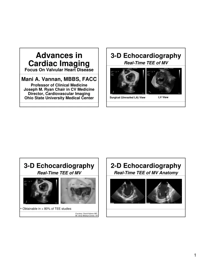
3-D Echocardiography
Real-Time TEE of MV
Courtesy: David Adams MD,
- Mt. Sinai Medical Center, NY
- Obtainable in > 80% of TEE studies
Surgical (Unroofed LA) View LV View