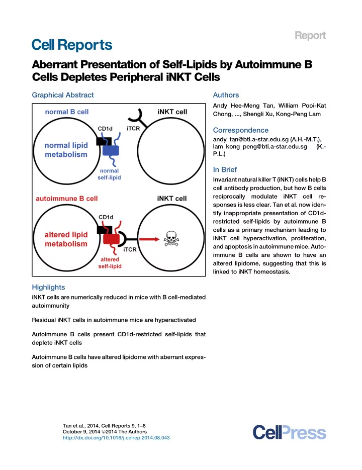

Report Aberrant Presentation of Self-Lipids by Autoimmune B Cells Depletes Peripheral iNKT Cells Graphical Abstract Authors Andy Hee-Meng Tan, William Pooi-Kat Chong, ..., Shengli Xu, Kong-Peng Lam Correspondence andy_tan@bti.a-star.edu.sg (A.H.-M.T.), lam_kong_peng@bti.a-star.edu.sg (K.- P.L.) In Brief Invariant natural killer T (iNKT) cells help B cell antibody production, but how B cells reciprocally modulate iNKT cell re- sponses is less clear. Tan et al. now iden- tify inappropriate presentation of CD1d- restricted self-lipids by autoimmune B cells as a primary mechanism leading to iNKT cell hyperactivation, proliferation, and apoptosis in autoimmune mice. Auto- immune B cells are shown to have an altered lipidome, suggesting that this is linked to iNKT homeostasis. Highlights iNKT cells are numerically reduced in mice with B cell-mediated autoimmunity Residual iNKT cells in autoimmune mice are hyperactivated Autoimmune B cells present CD1d-restricted self-lipids that deplete iNKT cells Autoimmune B cells have altered lipidome with aberrant expres- sion of certain lipids Tan et al., 2014, Cell Reports 9, 1–8 October 9, 2014 ª 2014 The Authors http://dx.doi.org/10.1016/j.celrep.2014.08.043
Please cite this article in press as: Tan et al., Aberrant Presentation of Self-Lipids by Autoimmune B Cells Depletes Peripheral iNKT Cells, Cell Reports (2014), http://dx.doi.org/10.1016/j.celrep.2014.08.043 Cell Reports Report Aberrant Presentation of Self-Lipids by Autoimmune B Cells Depletes Peripheral iNKT Cells Andy Hee-Meng Tan, 1,2, * William Pooi-Kat Chong, 3 Sze-Wai Ng, 2 Nurhidayah Basri, 3 Shengli Xu, 1,4 and Kong-Peng Lam 1,4,5,6, * 1 Immunology Group 2 Microarray Group 3 Metabolomics Group Bioprocessing Technology Institute, Agency for Science, Technology and Research, Singapore 138668, Singapore 4 Department of Physiology 5 Department of Microbiology 6 Department of Pediatrics Yong Loo Lin School of Medicine, National University of Singapore, Singapore 117599, Singapore *Correspondence: andy_tan@bti.a-star.edu.sg (A.H.-M.T.), lam_kong_peng@bti.a-star.edu.sg (K.-P.L.) http://dx.doi.org/10.1016/j.celrep.2014.08.043 This is an open access article under the CC BY-NC-ND license (http://creativecommons.org/licenses/by-nc-nd/3.0/). SUMMARY et al., 2011) bacteria. Recognition of foreign and self-lipids is critical to the immunomodulatory functions of iNKT cells Invariant natural killer T (iNKT) cells provide cognate (Godfrey and Kronenberg, 2004; Taniguchi et al., 2003), which help via CD1d to lipid antigen-presenting B cells for include the promotion of B cell antibody production, plasma cell generation, and memory B cell recall responses (Barral antibody production, but whether B cells reciprocally et al., 2008; Galli et al., 2007; Leadbetter et al., 2008; Tonti regulate iNKT cells remains largely unexplored. Here, et al., 2009). we found peripheral, but not thymic, iNKT cells to be How B cells in turn modulate iNKT cell responses is far less numerically reduced in autoimmune mice lacking Fas known. Human B cells capture exogenous lipid antigens using specifically in B cells. The residual iNKT cells were apolipoprotein E and low-density lipoprotein receptor, leading antigenically overstimulated, had altered cytokine to enhanced lipid presentation via CD1d to iNKT cells (Allan production, and manifested enhanced proliferation et al., 2009). Splenic marginal zone (MZ) B cells also trigger and apoptosis. B cell-specific ablation of CD1d iNKT cells to proliferate and produce interleukin-4 (IL-4) and ameliorated these iNKT defects, suggesting that inap- IL-13 (Zietara et al., 2011) and preferentially provide cytokine propriate presentation of CD1d-restricted self-lipids signals to dendritic cells for optimal iNKT activation (Bialecki by autoimmune B cell-depleted peripheral iNKT cells. et al., 2009). Recently, B cells from systemic lupus erythemato- CD1d + autoimmune B cells have reduced a -galactosi- sus (SLE) patients were shown to activate iNKT cells poorly compared with those from healthy donors and resulted in dase A expression and, as revealed by lipidomic reduced iNKT cell frequencies in their peripheral blood (Bosma profiling, altered lipidome with aberrant accumulation et al., 2012). A similar decrease in iNKT cells was observed in pa- of certain self-lipids and reduction of others. These tients with type 1 diabetes (Kukreja et al., 2002) and inflammatory findings unveil a critical link between autoimmunity, arthritis (Tudhope et al., 2010). However, it was not clear if iNKT B cell lipidome, and the maintenance of peripheral reduction resulted from genetic mutations affecting iNKT cells iNKT cells and highlight an essential homeostatic intrinsically or extrinsically via antigen-presenting cells or both. function of B cells beyond antibody production. Moreover, studies with human blood leukocytes might not reflect how B and iNKT cells interact in peripheral organs. INTRODUCTION Here, we found peripheral iNKT cells to be hyperactivated but numerically reduced in mice with FAS (CD95, Apo-1) ablated in B Invariant natural killer T (iNKT) cells are specialized T cells cells (Hao et al., 2008). Further analysis revealed autoimmune B bearing NK lineage receptors and T cell receptors (TCRs) cells have altered lipidome and disrupted iNKT cell homeostasis comprising an invariant V a -J a chain preferentially associated in a CD1d-dependent manner. with a limited set of V b chains that recognize glycolipid antigens presented by CD1d (Bendelac et al., 2007). These antigens RESULTS AND DISCUSSION include glycosphingolipids such as a -galactosylceramide ( a -GalCer) (Kawano et al., 1997) and isoglobotrihexosylceramide iNKT cells were reported to be numerically reduced in germline (Zhou et al., 2004) and glycolipids from Gram-negative (Kinjo MRL- lpr ( Fas mutant) mice and autoimmune patients (Bosma et al., 2005; Mattner et al., 2005) and Gram-positive (Kinjo et al., 2012; Yang et al., 2003), although it was not clear why Cell Reports 9 , 1–8, October 9, 2014 ª 2014 The Authors 1
Please cite this article in press as: Tan et al., Aberrant Presentation of Self-Lipids by Autoimmune B Cells Depletes Peripheral iNKT Cells, Cell Reports (2014), http://dx.doi.org/10.1016/j.celrep.2014.08.043 Figure 1. Reduced Peripheral iNKT Frequencies and Numbers in Autoimmune Fas f/f Cd19 Cre/+ Mice (A and B) Frequencies (A) and numbers (B) of iNKT cells in spleens, livers, bone marrows and thymi of 20-week-old Fas +/+ Cd19 Cre/+ and Fas f/f Cd19 Cre/+ mice as enumerated by flow cytometry. (C) Rearranged V a 14-J a 18 transcript levels in Fas +/+ Cd19 Cre/+ and Fas f/f Cd19 Cre/+ splenocytes as assessed by quantitative real-time PCR. Data in (A) are representative of more than four mice. Each symbol in (B) represents an individual mouse and small horizontal bars indicate the mean. Data in (C) are mean ± SEM of measurements obtained with four mice of each genotype. *p < 0.01; **p < 0.001; ns, not significant. See also Figure S1. they declined in these subjects. Here, we studied autoimmune iNKT cells. In contrast, iNKT cell frequencies and numbers in Fas f/f Cd19 Cre /+ mice lacking FAS in B cells and also found the thymi of Fas +/+ Cd19 Cre/+ and Fas f/f Cd19 Cre/+ mice were com- iNKT frequencies and numbers to be substantially reduced in parable. Thus, our data indicate that peripheral, but not thymic, iNKT cells are perturbed in autoimmune Fas f/f Cd19 Cre/+ mice. spleens, liver and bone marrows of 20-week-old mutants compared with age-matched Fas +/+ Cd19 Cre/+ controls (Figures To understand the cause of iNKT cell diminution in Fas f/f Cd19 Cre/+ mice, we examined their expression of costimulatory 1A and 1B). This defect was also apparent in young 8-week- old Fas f/f Cd19 Cre/+ mice (Figure S1). To exclude the possibility molecules and found higher expression of FAS, PD-1, ICOS, that the observed reduction in iNKT frequencies was due to cells OX-40, FASL, CD40L, PD-L1, and PD-L2 but normal CD25 and CD28, suggesting that Fas f/f Cd19 Cre/+ iNKT cells were hyperacti- internalizing their invariant TCRs, we assessed the levels of vated compared with Fas +/+ Cd19 Cre/+ counterparts (Figure 2A). In rearranged splenic V a 14 - J a 18 transcripts (normalized to C a ) by quantitative real-time PCR. We found them to be lower in addition, they have altered cytokine production as a greater frac- Fas f/f Cd19 Cre/+ compared with Fas +/+ Cd19 Cre/+ splenocytes tion (39.5%) of iNKT cells from Fas f/f Cd19 Cre/+ mice expressed (Figure 1C), confirming that mutant mice indeed had fewer interferon- g (IFN- g ) while a correspondingly lower fraction 2 Cell Reports 9 , 1–8, October 9, 2014 ª 2014 The Authors
Recommend
More recommend